Menu
Free Consultation
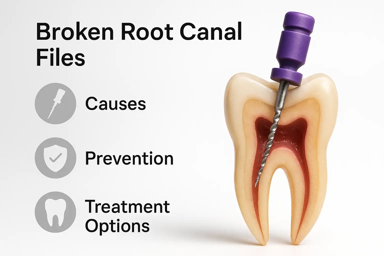
Endodontic (root canal) treatment is one of the most common procedures in dentistry. However, during this procedure, the instruments used to clean and shape the root canal system – called files – may fracture inside the canal. This “broken endodontic file” situation can be challenging for both the dentist and the patient.
In this comprehensive guide, we will examine why file separation occurs, its risk factors, prevention methods, and treatment options.
A root canal file is a metal instrument used to shape and clean the inside of the root canal. These files are typically made of stainless steel or nickel-titanium (NiTi) alloy.
A broken file refers to the situation where a file fractures during treatment, leaving a portion of it inside the root canal. This fragment may block the canal and prevent complete cleaning or filling.
In many cases, file separation occurs in the apical third of the canal (near the root tip) or in curved areas, which are anatomically narrower and harder to access, making removal more complex.
Additionally, improper working length determination can cause the file to bind apically, increasing fracture risk.
Most of the time, the dentist notices the instrument separation during the procedure.
However, some patients may present later with pain, swelling, or persistent discomfort.
Accurate diagnosis is crucial to plan the next steps and select the most suitable treatment approach.
A separated file can compromise the success of root canal treatment.
However, in many cases, the tooth can still be treated successfully and remain functional for many years, especially if the issue is managed promptly.
Files should be carefully examined visually and under magnification before each use. Any distortion or unwinding should be a reason for disposal.
Maintain continuous irrigation with sodium hypochlorite, EDTA, or other recommended solutions to keep the canal moist and reduce friction.
Establish a smooth, reproducible glide path with hand files before using rotary instruments. This reduces torsional stress and lowers fracture risk.
Experienced clinicians are better able to anticipate challenging canals and select the correct file sequence accordingly.
Additional precautions include radiographic planning, use of crown-down or step-back techniques, and avoiding excessive apical pressure during preparation.
The management of a separated instrument depends on its location, the length of the fragment, and the condition of the canal.
Whenever possible, the fragment is removed.
If removal is not feasible, the canal may be negotiated around the fragment, allowing cleaning and filling of the remainder of the canal.
In some cases, the file is left in place and the canal is filled as best as possible. The tooth is then monitored radiographically over time.
If the fragment is located in the apical region and cannot be removed non-surgically, apicoectomy and retrograde filling may be performed.
Modern dentistry relies heavily on dental operating microscopes and CBCT imaging for successful management of separated instruments.
Success depends on multiple factors, including the fragment’s location, operator experience, and available technology.
The use of operating microscopes and ultrasonics has significantly improved success rates.
Prompt intervention and careful planning increase the chances of saving the tooth.
Broken endodontic files are one of the most complex complications in root canal treatment, but thanks to modern techniques, they are no longer a hopeless scenario.
Encountering a broken file does not necessarily mean losing the tooth. With the right approach and technology, the tooth can often be preserved for many years, maintaining function and aesthetics.
Does a broken file mean I will lose my tooth?
No. In many cases, the file can be retrieved or bypassed, allowing the tooth to be treated and kept functional.
Can a broken file cause pain?
It may cause pain if it leads to persistent infection. Timely follow-up is essential.
How much does broken file treatment cost?
The use of a dental microscope and advanced equipment may increase the cost, but it is often worth it to save the tooth.
What happens if the file is not removed?
Sometimes the fragment is left in place and the tooth is monitored. If infection develops later, surgical intervention may be required.
How long does it take to remove a broken file?
Depending on the case complexity, it may take a single appointment or several visits. Microscope assistance usually shortens the procedure time.