Menu
Free Consultation
Root canal treatment is one of the most delicate and important procedures in dentistry. Its purpose is to remove infected or damaged tissue from the tooth, disinfect the canals, and seal them to preserve the tooth. However, during this procedure, endodontic files – the small metal instruments used to clean and shape the canals – can sometimes fracture and remain inside the tooth.
This is a situation that can worry both patients and dentists and raises the big question: Can a broken file be removed?
The answer depends on several factors, including the file’s location, the tooth’s anatomy, and the technology available. In this detailed guide, we’ll explore everything you need to know about separated instruments, their causes, removal techniques, prognosis, and prevention.
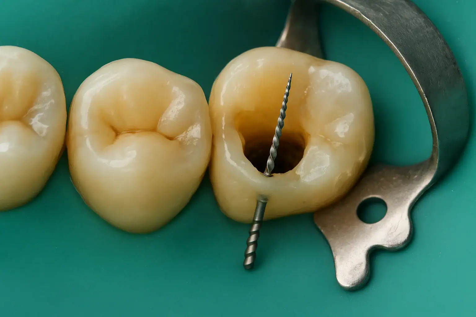
An endodontic file is a fine metal instrument used to shape and clean the root canal system. These files are usually made of stainless steel or nickel-titanium (NiTi) alloy.
A separated file is a piece of that instrument that fractures during the procedure and stays lodged inside the canal.
Why it matters:
Fortunately, encountering a separated instrument does not necessarily mean the tooth must be extracted. With the right approach, it can often be saved.
NiTi files are flexible, but repeated bending in curved canals leads to cyclic fatigue, eventually causing fracture.
A dry or poorly lubricated canal increases friction and torsional stress, contributing to file separation.
Most dentists notice the separation during the procedure itself, but in some cases it is discovered later, when symptoms such as pain, swelling, or persistent infection appear.
Diagnostic tools include:
In many cases, yes – but the success depends on several key factors:
CBCT provides a three-dimensional view, allowing precise assessment of:
The operating microscope offers high magnification and illumination, enabling the clinician to work conservatively and with minimal risk to tooth structure.
In a clinical case, a NiTi rotary file fractured in the mesiobuccal canal of a maxillary first molar. CBCT imaging identified the fragment’s position.
Using an operating microscope and ultrasonic tips, the dentist successfully retrieved the fragment, disinfected the canal, and completed obturation.
At the one-year follow-up, radiographs showed complete healing of the periapical lesion – confirming that even difficult cases can have excellent outcomes with the right approach.
Hearing that an instrument broke during treatment can be stressful for patients.
A good clinician should:
Experienced endodontists use magnification and precision instruments to minimize these risks.
After retrieval or bypassing of the fragment:
With modern techniques and equipment, success rates range between 70–90%, depending on fragment location and case complexity.
Even if the fragment cannot be removed, bypassing and proper sealing can allow the tooth to function for many years without symptoms.
File separation during root canal treatment is a challenging complication, but with today’s advanced technology, it is highly manageable.
Bottom line: A broken instrument does not necessarily mean the tooth will be lost. With early intervention, proper planning, and expert care, the tooth can often be preserved for many years.
Can a broken file always be removed?
No. In some cases, it cannot be removed, but bypassing it is possible to complete treatment.
Will the tooth need to be extracted if the file stays inside?
Not necessarily. The tooth can often remain functional under close monitoring.
Is the removal procedure painful?
No – it is done under local anesthesia, making it painless for the patient.
What is the success rate?
With proper equipment and expertise, success rates can be as high as 70–90%.