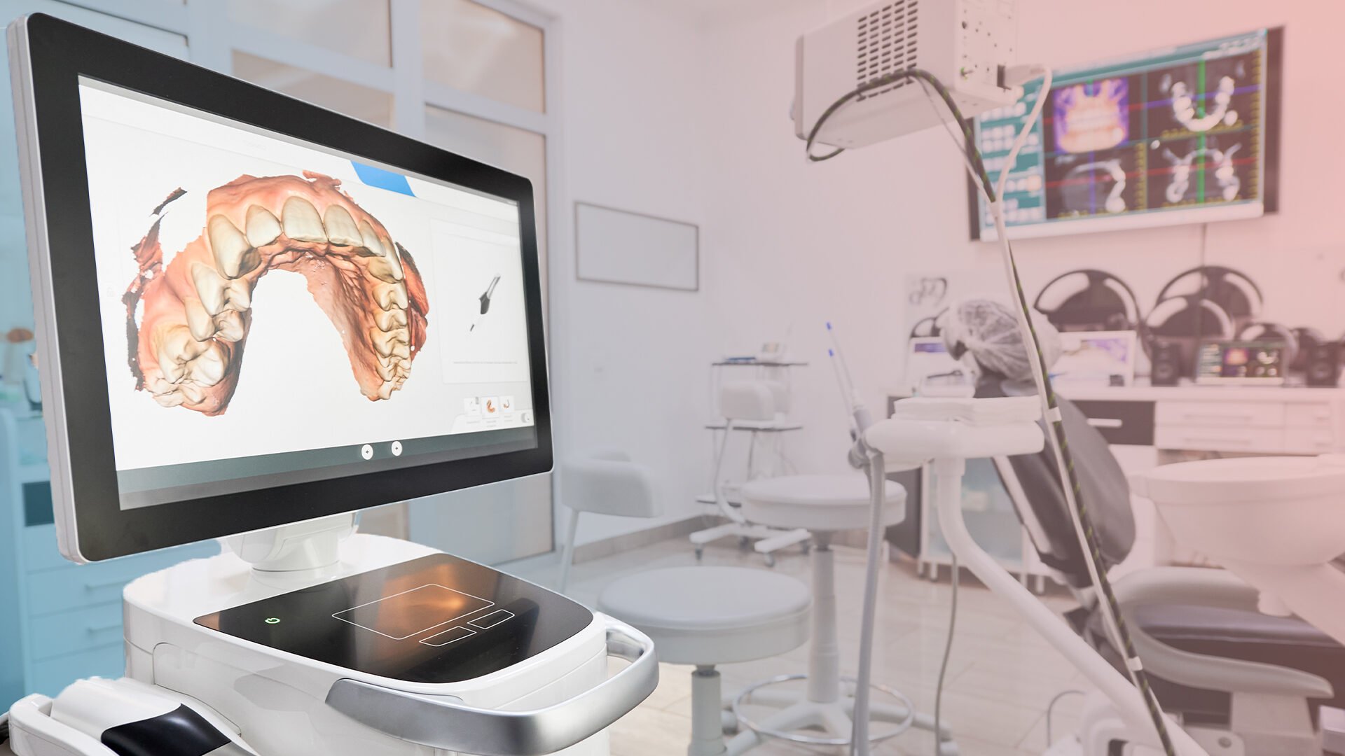
Digital dentistry is the delivery of dental care through computer-aided systems, in which the impression, the diagnosis, the treatment plan and the fabrication all run on a shared three-dimensional data set instead of trays, stone models and manual waxing. Its accuracy is measurable rather than absolute: in an in vivo study of 11 participants scanned with three intraoral scanners, median trueness was 38 to 47 micrometres for short-span scans and 147 to 433 micrometres for complete-arch scans (PubMed 33902891). The working tools are intraoral scanners, CAD/CAM design and milling, 3D printing and digital radiography, and the practical gain is fewer appointments and same-visit restorations.
- 01Digital dentistry replaces the analogue chain of tray impression, stone model and hand fabrication with a scan, design and manufacture chain built on one 3D file.
- 02Scanner accuracy depends on the span being scanned: median in vivo trueness was 38 to 47 micrometres for short-span scans against 147 to 433 micrometres for complete-arch scans, across three devices and 11 participants (PubMed 33902891).
- 03In the same study, extraoral scanning of the model was more precise and truer than intraoral scanning for almost every comparison, and the authors concluded that intraoral conditions reduced scanning accuracy (PubMed 33902891).
- 04Full-arch implant scanning is a separate measurement problem: resultant trueness was 41 plus or minus 11 micrometres with the control protocol, 49 plus or minus 22 with dental floss and 55 plus or minus 8 with an acrylic splint (PubMed 36481688).
- 05CAD/CAM shortens the laboratory step rather than the clinical one: crowns, onlays, veneers and temporary restorations can be designed and milled in hours, which removes shipping and model pouring from the timeline.
What Digital Dentistry Replaces in the Traditional Workflow
Digital dentistry is the delivery of dental care through computer-aided systems, where the impression, the diagnosis, the treatment plan and the fabrication all run on one shared three-dimensional data set. In the traditional chain, a tray filled with silicone takes the impression, a technician pours a stone model, and the restoration is waxed and cast by hand. Each of those steps adds time and a small opportunity for distortion. In the digital chain, an intraoral scanner records the teeth as a 3D file, design software builds the restoration on that file, and a milling unit or a 3D printer produces it.
The technology did not arrive at once. CAD/CAM systems were first used for fixed prosthesis design and spread gradually into general practice. The toolkit in a modern clinic now includes digital impression systems, three-dimensional printers, diagnostic software that assists with reading images, digital radiography and cone beam tomography, and in-clinic production units that take over part of the laboratory work.
The practical gain for the patient is shorter timelines and fewer visits. Prosthesis work that could take weeks with the traditional chain can, in suitable cases, be completed in hours, because the model pouring and shipping stages disappear. The gain has limits, and those limits are measurable. Scanning accuracy changes with the span of the arch, full-arch implant cases need specific protocols, and complex restorations still require healing time and laboratory finishing.
How Accurate Intraoral Scanners Are
An intraoral scanner is accurate enough for short-span work such as single crowns, and noticeably less accurate across a complete arch. The scanner replaces the impression tray and silicone, which many patients find uncomfortable, and it is especially helpful for people with a strong gag reflex. The inside of the mouth is captured in a series of images that the software stitches into a 3D model, and the dentist can review and record it while the patient is still in the chair.
Accuracy has been measured in the mouth rather than on a bench. In an in vivo study, an experimental appliance was fabricated for 11 participants and scanned with 3 different intraoral scanners and a reference scanner (PubMed 33902891). Median trueness of intraoral short-span scans was 38 to 47 micrometres, while intraoral complete-arch scans reached 147 to 433 micrometres (PubMed 33902891). Precision followed the same pattern: 23 to 43 micrometres for short-span intraoral scans against 80 to 198 micrometres for complete arches (PubMed 33902891).
The same study found that extraoral scans of the appliance were more precise and truer than intraoral scans, with one exception among the complete-arch comparisons, and the authors concluded that intraoral conditions reduce scanner accuracy (PubMed 33902891). For this reason, claims that a scanner works to a thousandth of a millimetre do not match the clinical data. A single crown and a full-arch restoration are different scanning tasks, and the clinical plan should reflect that difference.
Full-Arch Implant Scanning and Protocol Choice
Scanning a full arch of implants is used clinically, but its accuracy depends on how the scan is taken. In implant work the scanner records scan bodies screwed into the implants, and the software converts their position into the position of each implant. Small errors across a long span can add up, and a full-arch bridge must fit several implants at once.
An in vivo study on edentulous upper jaws measured this directly. Six scan bodies were mounted on maxillary implants in five subjects, and each subject was scanned with three different protocols (PubMed 36481688). Resultant trueness was 41 plus or minus 11 micrometres with the control protocol, 49 plus or minus 22 with a dental floss aid and 55 plus or minus 8 with an acrylic splint (PubMed 36481688). The authors found the control protocol the most accurate and noted that a completed scan does not necessarily equate to an accurate scan (PubMed 36481688).
The study was small and described as a pilot, and the authors state that other scanners may behave differently in vivo. For the patient, the lesson is practical. Digital planning in implant treatment analyses the angle at which the implant will be placed, its depth and the density of the jawbone in advance, and surgical guides printed from that plan help transfer it to the mouth. The final restoration still depends on a careful scanning protocol, a trial fit where needed and checking the result against the plan before the bridge is fixed.
CAD/CAM Design and 3D Printing in Everyday Care
CAD/CAM shortens the laboratory stage of treatment; it does not shorten the clinical stages that depend on healing. CAD stands for computer-aided design and CAM for computer-aided manufacturing. The restoration is designed on screen from the scan file and then milled from a ceramic or composite block, or printed. Crowns, bridges, veneers, inlays, onlays and temporary restorations can all be produced this way.
When a clinic has chairside design and milling capacity, a single crown can be prepared, scanned, designed and milled in the same session. The temporary crown stage and the second appointment can then be skipped. Complex cases, implant restorations and full-arch work still run on a longer schedule, because the tissue has to heal and the restoration often needs laboratory finishing, staining and glazing. Digital production also reduces some manual steps, which lowers the chance of certain handling errors, but a digital restoration is still checked for fit, contact and bite before it is cemented, and adjustments or remakes are sometimes needed.
3D printers extend the same idea to other appliances. Night guards, temporary teeth, orthodontic appliances and surgical guides can be produced directly from the digital model. This shortens the time between planning and delivery and keeps a digital copy of the design, so a lost or broken appliance can be reproduced from the file.
Digital Radiography, Radiation Dose and Image Reading
Digital X-rays give instant images and can lower the dose a patient receives, and the size of the dose depends on the type of examination and the equipment. Digital images appear on screen within seconds, are stored directly in the patient record, and can be enlarged and filtered to examine details.
Dose has been measured with a head phantom that mimics human tissue. Effective dose was 14.2 microsieverts for one digital panoramic unit and 24.3 microsieverts for another, against 388 microsieverts for a full set of intraoral films on D-speed film with round collimation (PubMed 18762634). The authors list digital receptors or faster film, rectangular collimation and radiographic selection criteria as ways to reduce patient dose substantively (PubMed 18762634). A panoramic film therefore carries a small dose, and the decision to take any X-ray still rests on a clinical reason.
Software that assists image reading is a newer layer. Such programmes scan radiographs and highlight areas that may indicate caries, root canal needs, gum disease or changes in bone density. They can speed up the review and help the dentist notice findings, but they do not make the diagnosis on their own. The dentist compares the highlighted areas with the clinical examination, and the treatment decision remains a clinical one.
Digital Planning Across Dental Specialties
Digital planning is now used in most branches of dentistry, and its role differs from one field to another. In aesthetic work, a smile simulation is prepared on screen by taking into account the person's facial structure, smile line and the ratio between tooth and lip. In smile design, this lets the patient see a proposed result before any tooth is prepared and discuss changes with the dentist while they are still easy to make.
In orthodontics, clear aligner treatment is planned entirely in software. The patient can follow the planned tooth movement in 3D before the first aligner is worn, which helps set expectations and can support motivation during treatment. The plan is a forecast, and the actual movement is checked at follow-up visits and corrected where it lags behind.
In paediatric dentistry, replacing tray impressions with a scan removes a step that many children find unpleasant, and some clinics use playful on-screen tools to help children feel at ease. In endodontics and restorative care, electronic canal length measurement supports root canal treatment, and CAD-designed fillings such as inlays and onlays can be milled from durable ceramic or composite blocks.
Education, Patient Records and Data Security
Digital tools have changed how dentists train and how clinics store information, and both changes bring responsibilities. Dental students now practise on simulation software and digital patient models, planning restorations and rehearsing procedures before they treat real patients.
Looking ahead, research is exploring robotic assistance, remotely controlled devices, more automated prosthesis production and smaller, mobile intraoral scanners. These are directions of development, and their place in routine care will depend on the clinical evidence that follows them.
Because scans, X-rays and treatment plans are stored as digital files, data security is part of patient care. Clinics protect records with SSL certified systems, encrypted access to data, cloud backup and transaction approvals with an electronic signature. In Turkey, archiving has to comply with KVKK, the national personal data protection law, which sets rules on who can access health data and how long it is kept.
For the patient, the questions worth asking are simple. Which steps of my treatment are digital, and which still go through a laboratory? Will my scan be single-span or full-arch, and how will the fit be checked? Which X-rays are planned, and why? How is my data stored? Clear answers to these questions show how the digital workflow will be used in your own treatment.
Let's plan the right treatment together.
Free Assessment→- 1.Kernen F, Schlager S, Seidel Alvarez V, Mehrhof J, Vach K, Kohal R, et al. Accuracy of intraoral scans: An in vivo study of different scanning devices. J Prosthet Dent 2022;128:1303-1309. PubMed 33902891
- 2.Nedelcu R, Olsson P, Thulin M, Nyström I, Thor A. In vivo trueness and precision of full-arch implant scans using intraoral scanners with three different acquisition protocols. J Dent 2023;128:104308. PubMed 36481688
- 3.Ludlow JB, Davies-Ludlow LE, White SC. Patient risk related to common dental radiographic examinations: the impact of 2007 International Commission on Radiological Protection recommendations regarding dose calculation. J Am Dent Assoc. 2008;139(9):1237-43. PubMed 18762634