Menu
Free Consultation
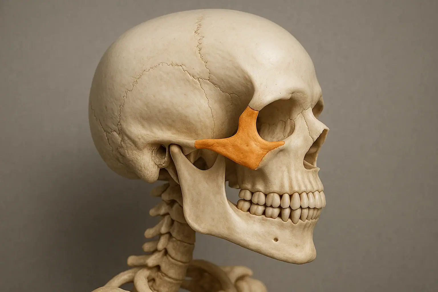
The human skull is a structural masterpiece, and among its most recognizable features is the zygomatic bone, also commonly known as the cheekbone. Not only does it define the contours of the midface, but it also supports essential functions including protection of the eye socket, attachment for facial muscles, and articulation with surrounding cranial structures.
In this comprehensive guide, we’ll dive into everything you need to know about the zygomatic bone—its anatomy, articulations, development, clinical relevance, and aesthetic importance. Whether you're studying anatomy, pursuing dental or medical education, or simply curious about facial bones, this article provides the complete picture.
The zygomatic bone is a paired, irregular facial bone that gives shape to the cheeks and forms part of the lateral wall and floor of the orbit (eye socket). It is often referred to as:
It plays a vital role in both form and function by contributing to facial aesthetics and serving as a structural foundation for surrounding tissues.
The zygomatic bones are situated on each side of the face and are easily palpable just below and to the side of the eyes.
Anatomical Position:
These bones give the cheeks their prominence and serve as lateral buttresses for the midface.
Each zygomatic bone has several projections (or processes) that articulate with neighboring bones, contributing to the structural complexity of the skull.
The zygomatic bone forms joints with four bones:
| Bone | Connected Process |
|---|---|
| Frontal bone | Frontal process of zygomatic bone |
| Maxilla | Maxillary process |
| Temporal bone | Temporal process & zygomatic process |
| Sphenoid bone | Orbital surface |
These articulations support the orbit, cheek, and jaw structures and create stability across the midface.
The zygomatic bones are essential for both aesthetic and functional reasons:
Several muscles responsible for facial expression and mastication originate or insert on the zygomatic bone:
The zygomatic arch is formed by the union of:
It serves to:
A common facial injury involving:
Symptoms include:
Different ethnic populations display unique zygomatic morphologies, which can impact both facial aesthetics and surgical planning.
| Population | Zygomatic Characteristics |
|---|---|
| East Asian | Highly prominent, laterally flared |
| European | Balanced projection, moderate prominence |
| African | Broad, robust, and anteriorly angled |
It is a paired facial bone also known as the cheekbone, forming the prominence of the cheek and part of the eye socket.
A projection that connects with the zygomatic process of the temporal bone to form the zygomatic arch.
On the upper lateral side of the face, between the maxilla and the temporal bone, beneath the eye.
A projection of the temporal bone that articulates with the zygomatic bone.
Mild, non-displaced fractures may heal on their own, but many require surgical intervention for function and facial symmetry.
Zygomatic implants are anchored into the bone when maxillary bone mass is insufficient for traditional implants.
| Aspect | Details |
|---|---|
| Common Names | Zygoma, cheekbone, malar bone |
| Location | Lateral midface, below the eye socket |
| Processes Involved | Temporal, maxillary, frontal, orbital |
| Articulated Bones | Maxilla, temporal, sphenoid, frontal |
| Related Muscles | Zygomaticus major/minor, masseter, temporalis |
| Clinical Relevance | Fractures, implants, cosmetic surgery, facial reconstruction |
| Diagnostic Tools | CT, CBCT, X-ray, MRI |
| Development Origin | Neural crest, ossifies during fetal development |
| Anthropological Significance | Used in forensic identification and craniofacial classification |
The zygomatic bone is more than just the prominent curve of your cheek—it’s a crucial architectural, functional, and aesthetic cornerstone of the human face. It interacts with multiple bones and muscles, affects dental and ophthalmologic outcomes, and holds a prominent role in cosmetic, reconstructive, and forensic practices.
Whether you're a healthcare professional, anatomy student, or cosmetic surgeon, understanding the zygomatic bone, including its temporal process, zygomatic arch, and clinical implications, provides essential insight into facial harmony, structure, and health.