
Micro Endodontics
Micro endodontics is root canal treatment under a surgical microscope at high magnification, used for calcified canals, missed canals and retreatment.
- Surgical microscope
- Retreatment cases
- Complex anatomy management
First consultation free · same-day reply · no price pressure
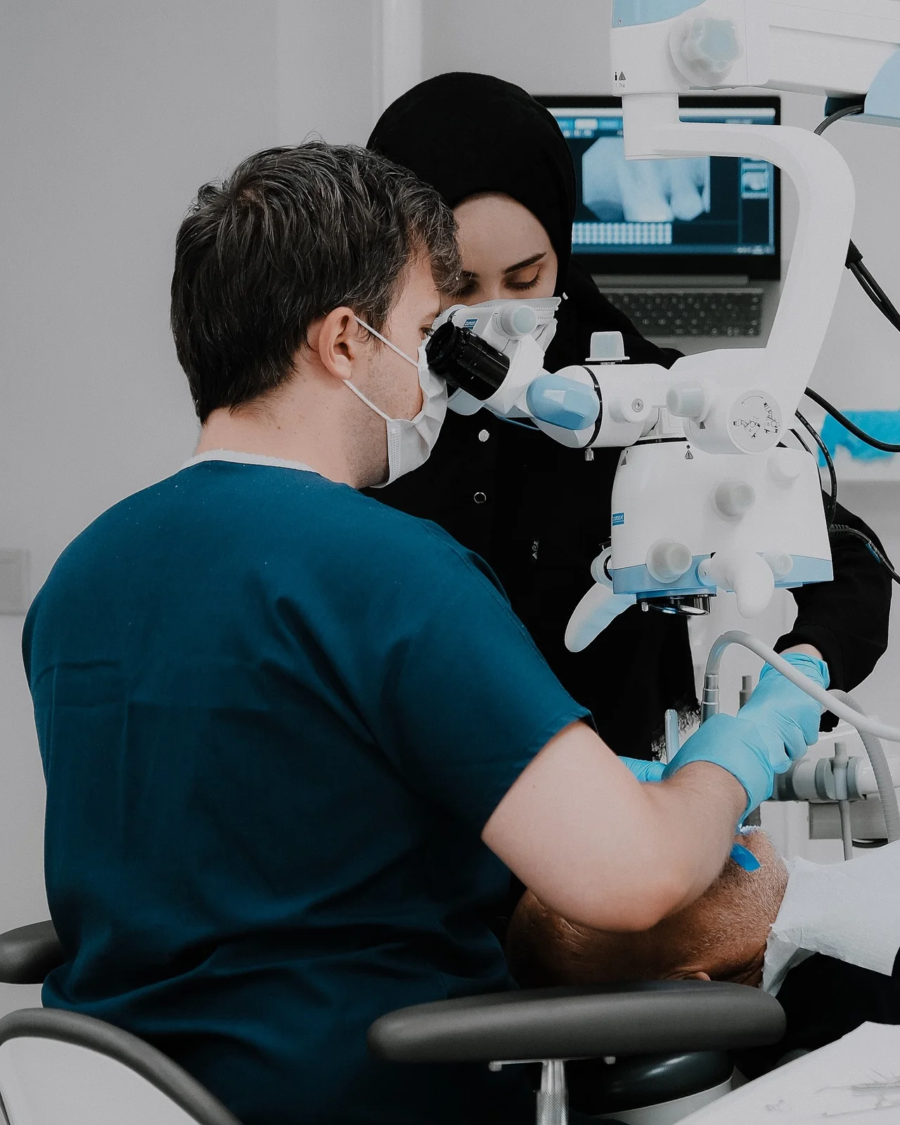
Micro endodontics is root canal treatment carried out under high magnification with a surgical operating microscope or endoscope, used for calcified canals, missed canals and retreatment of previously treated teeth. In a meta-analysis of 21 studies of root-end surgery, the weighted pooled success rate was 59% for traditional technique (12 studies, n=925) against 94% for endodontic microsurgery (9 studies, n=699), a statistically significant difference (P < .0005; PubMed 20951283). Part 2 of the same series compared operators using identical micro-instruments: 88% with loupes or no visual aid against 94% with a microscope or endoscope (PubMed 22152611).
- 01Most of the gain comes from micro-instruments and the remainder from magnification, which is worth knowing before paying a premium for the word microscope. The same series reports traditional root-end surgery at 59% against microsurgery at 94% (relative success 1.58), then micro-instruments with loupes at 88% against microscope or endoscope at 94% (relative success 1.07). The second difference was significant for molars (n=193, P = .011) but not for premolars or anterior teeth (PubMed 20951283, 22152611).
- 02Missed canals are a measured anatomical problem. A meta-analysis of 26 cone beam computed tomography studies found the second mesiobuccal canal present in 69.6% of 15,285 maxillary first molars (95% CI 64.5-74.8%) and 39.0% of 8,641 second molars (PubMed 31735252).
- 03Working without magnification is a clear disadvantage, but the microscope did not beat loupes at finding canals. Across 312 clinical cases, the MB2 canal was located in 57.4% with a microscope, 55.3% with loupes and 18.2% with no magnification; for maxillary first molars alone the figures were 71.1%, 62.5% and 17.2%, with no significant difference between microscope and loupes (PubMed 12043874).
- 04Retreatment has a published expected outcome you can hold a plan to. A systematic review of 17 studies put the pooled weighted success rate of secondary root canal treatment at 76.7% judged by complete healing (95% CI 73.6-89.6%), with pre-operative periapical lesion, apical extent of the root filling and quality of the coronal restoration as significant prognostic factors (PubMed 19133093).
- 05Micro endodontics names an imaging and lighting setup. The Setzer comparisons are built on exactly that premise: both groups used the same micro-instruments and the same root-end filling materials, and magnification was the only variable (PubMed 22152611).
/ Micro Endodontics Definition
Infection and/or necrosis of the living tissue inside the tooth requires root canal treatment. The tissues that keep the tooth in the mouth are actually healthy gum and jawbone tissues, not the vascular and nerve bundle inside the tooth. Therefore, with a successful root canal treatment, it may be possible to keep the tooth in the mouth for many years. On the other hand, the critical role of endodontic preventive dentistry should not be overlooked, both in terms of treating infection and preventing other treatment needs that may arise due to extraction.
Micro endodontics, also known as microscopic endodontics, is an advanced and modern method of root canal treatment using a microscope. Micro endodontics is related to endodontics, a specialized branch of dentistry. Endodontics is a branch that treats the pulp, the soft tissue inside the teeth, and the roots of the teeth. Micro-endodontics is a method that allows these treatment methods to be applied in a more precise and detailed way.
Microendodontics refers to an approach that involves the use of a microscope. During endodontic procedures, microscopes are used to better visualize and treat the small canals inside the teeth. This method gives dentists a better view and allows procedures to be performed more precisely.
Microendodontics involves performing endodontic procedures such as pulp treatment, root canal treatment, apical resection under a microscope. In this way, more successful results are achieved and patient comfort is increased. Microendodontics is considered a significant advancement towards improving the quality of endodontic treatments with the use of technological advances in dentistry.
| At a glance | |
|---|---|
| Duration | 1-2 sessions |
| Sessions | 2 |
| Warranty | 2 years |
| Materials Used | Surgical microscope · Ultrasonic tips |
/ How is Micro Endodontics Applied?
Microendodontics is a sub-branch of endodontics that enables treatments performed on the internal structure of the teeth to be performed more precisely and effectively, supported by microscopic imaging and specialized equipment. This innovative approach provides dentists with a clearer view and improves the treatment process. Thus, it may be possible to increase patient comfort and treatment success.
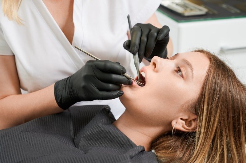
Microendodontics is performed in a similar way to traditional endodontic procedures. However, it is performed in a more detailed and precise manner with the use of microscopic imaging and specialized instruments. Microendodontic treatment begins with a patient examination and digital imaging techniques. The dentist diagnoses the problem by examining the internal structure of the tooth in detail.
Before the procedure begins, the dentist will administer an appropriate local anesthetic for the patient. A rubber dam is then placed around the tooth to keep the procedure area dry and provide a suitable environment for microendodontics.
Any decayed or damaged tissue on the tooth is removed. The dentist accesses the internal structure of the tooth using the microendodontic microscope. Then, the process of removing infected or damaged tissues in the pulp area begins.
Under the microendodontic microscope, the root canals are carefully enlarged and shaped. The next step is to fill the canals with special endodontic filling materials. An impermeable barrier is created to prevent leakage. Once root canal treatment is complete, an appropriate filling or crown is placed to restore the tooth's structure. Thus, the function and aesthetics of the tooth can be restored.
Treatment Process
- 01Examination: Diagnosis with digital imaging, then local anaesthesia and rubber-dam isolation.
- 02Cleaning & Shaping: Under the microscope the root canals are carefully enlarged and shaped.
- 03Filling & Restoration: Canals are filled with special endodontic materials and the tooth is restored.
/ Is Microendodontics Treatment Painless?
Root canal treatment can be defined as the process of removing, cleaning and refilling the damaged tissues inside the tooth layer. The pulp layer can be damaged by tooth decay. Although there is nothing visible in painful teeth, if there is a lesion at the root tip, root canal treatment is required. A good root canal treatment is very important for maintaining dental health.
Microendodontic treatment, like other endodontic procedures, is usually associated with minimal pain or discomfort. However, each patient is different and experiences may vary due to the complexity of the treatment, the patient's pain threshold and other factors.
Microendodontics is performed more precisely under a microscope. This also allows dentists to carry out treatment processes more accurately. Thus, less tissue is damaged during the procedure and a more effective treatment can be performed. Furthermore, microendodontic techniques and modern anesthesia methods are designed to ensure that patients have a comfortable and pain-free experience during the procedure.
Before microendodontics, the dentist explains in detail to the patient what steps will be followed and what measures will be taken to minimize any pain. Also, if the patient feels any discomfort during the procedure, they should inform the dentist. In this way, necessary precautions can be taken.
/ Root Canal Treatment Under the Microscope
Root canal treatment under the microscope provides more successful results than the classical root canal treatment method. Because the roots and pulp base, which cannot be seen with the naked eye, can be easily seen thanks to the microscopic endodontic method. By utilizing the magnification feature of the microscope, root canals are filled better and more effective treatments can be performed.
There are many cases where root canal treatment is very difficult to perform due to complex tooth anatomy. Microscopes are extremely useful tools in endodontic procedures. Micro endodontics offers many benefits in dentistry procedures in this field, such as greater accuracy, better visualization, treatment of complex cases, better diagnosis and many more. It is also considered an excellent treatment option for teeth that have previously failed root canal treatments for various reasons.
Severe tooth decay or other trauma creates open doors for bacteria to reach the interior of the tooth. When the nerves and blood vessels in the tooth are damaged by bacterial contamination, in most cases the tooth becomes painful. The aim of root canal procedures (endodontic treatments) is to save the painful tooth by removing the pulp tissue, which consists of nerves and blood vessels, from the canals inside the tooth. The canals are then cleaned, shaped and filled with a special material. The tooth is usually restored with a crown. In some cases, surgical removal of the root tip is required.
As in most teeth, a canal containing nerves and blood vessels runs from the center of the crown of the tooth through the root and into the jaw. Near the tip of the root, it divides into many smaller canals. These small branches of the canal must be completely cleaned and sealed to stop pain and ensure an effective and long-lasting result. These small canals are much easier to see under a microscope.
/ Advantages of Microscope Imaging in Root Canal Treatment
- Root canal treatment is the process of cleaning and filling the infected or damaged tissues and nerves inside the teeth. This treatment saves the tooth and prevents pain and tooth loss. In traditional root canal treatments, dentists usually only work with the naked eye or x-ray images. The use of microscopes, which has come to the fore in recent years, has made a significant progress in the field of root canal treatment.
- Root canal treatment under a microscope provides the dentist with a much more detailed view. Small root canals and anatomical structures that cannot be seen in traditional methods can be seen clearly under the microscope. This allows the dentist to work more accurately and effectively during treatment.
- Root canal treatment under a microscope makes the procedure more precise. The microscope allows the dentist to gain better access to the root canals and remove infected tissues more effectively. This increases the success of the treatment and reduces the risk of complications.
/ Microsurgery and Tissue Protection with Microscope Use
- The use of a microscope makes it possible to apply microsurgical techniques. This contributes to the surgical removal of infected tissues at the root tip and to the reconstruction of the root tip. At the same time, the success of the treatment is increased and patient comfort is optimized.
- Root canal treatment performed under a microscope results in less tissue damage. When traditional methods are used, a larger area may be affected in order for the dentist to clean the infected tissues. However, under the microscope, it is possible to work in a less invasive way by directly targeting the infected tissues.
- Root canal treatment under the microscope usually has a higher success rate. More detailed imaging, a more precise procedure and the possibility of microsurgery are considered to be the elements that enable the treatment to be performed more effectively. As a result, both the success of the treatment and the patient satisfaction rate can be increased.
In conclusion, the use of a microscope is a very important advance in the field of root canal treatment. With its advantages such as more detailed imaging, precise processing, microsurgery and high success rate, root canal treatment under the microscope raises the standards in dentistry.
- 1.Setzer FC, Shah SB, Kohli MR, Karabucak B, Kim S. Outcome of endodontic surgery: a meta-analysis of the literature--part 1: Comparison of traditional root-end surgery and endodontic microsurgery. J Endod 2010;36:1757-65. PubMed 20951283
- 2.Setzer FC, Kohli MR, Shah SB, Karabucak B, Kim S. Outcome of endodontic surgery: a meta-analysis of the literature--Part 2: Comparison of endodontic microsurgical techniques with and without the use of higher magnification. J Endod 2012;38:1-10. PubMed 22152611
- 3.Martins JNR, Marques D, Silva EJNL, Caramês J, Mata A, Versiani MA. Second mesiobuccal root canal in maxillary molars-A systematic review and meta-analysis of prevalence studies using cone beam computed tomography. Arch Oral Biol 2020;113:104589. PubMed 31735252
- 4.Buhrley LJ, Barrows MJ, BeGole EA, Wenckus CS. Effect of magnification on locating the MB2 canal in maxillary molars. J Endod 2002;28:324-7. PubMed 12043874
- 5.Ng YL, Mann V, Gulabivala K. Outcome of secondary root canal treatment: a systematic review of the literature. Int Endod J 2008;41:1026-46. PubMed 19133093

Who is this treatment for?
- Infection and/or necrosis of the living tissue inside the tooth
- A lesion at the root tip of a painful tooth
- Bacteria reaching the inside of the tooth after severe decay or trauma
- Teeth with persistent symptoms after previous root canal treatment
- Calcified canals
- Multi-rooted and anatomically complex teeth
- A broken instrument inside the root canal
Doctors specialising in this treatment




Frequently asked
01How does micro-endodontics differ from standard root canal treatment?+
A dental microscope provides x8-x25 magnification, enabling the detection and treatment of fine canals, broken instruments, and missed apices that are invisible with the naked eye.
02When is micro-endodontics recommended?+
Previously root-canal-treated teeth with persistent symptoms, calcified canals, and multi-rooted or anatomically complex teeth are the primary indications. Micro-endodontics is a form of root canal treatment carried out under a surgical microscope.
Endodontics
Endodontic Surgery
Endodontic surgery removes the last few millimetres of a root and seals the canal end if infection persists after a root canal and retreatment is impossible.
Explore→02Root Canal Treatment
Root canal treatment removes infected or devitalised pulp from inside the tooth, then cleans, shapes and fills the canals with gutta-percha to keep the tooth.
Explore→Speak with your doctor about this treatment.
Not sure which treatment fits? Tell us what’s wrong →