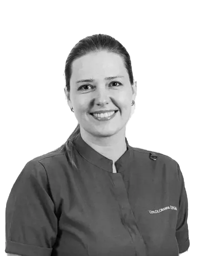
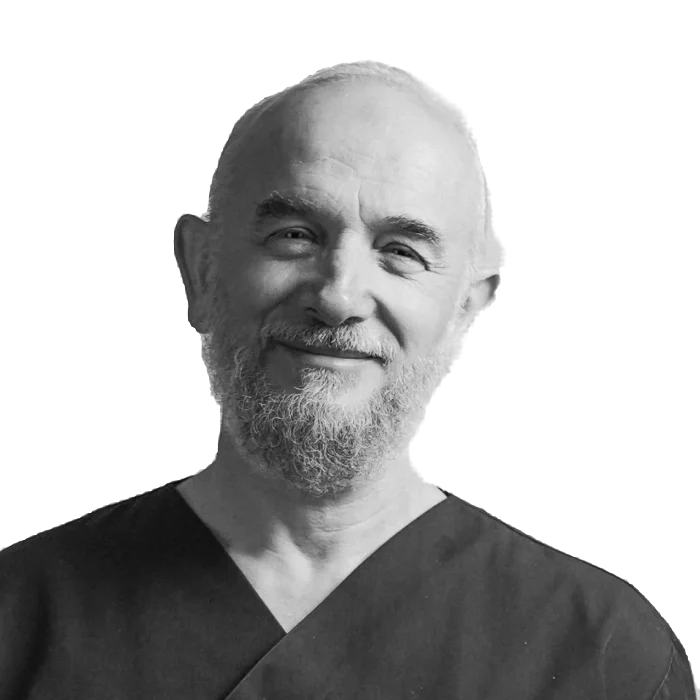
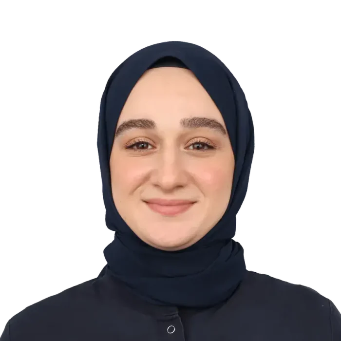
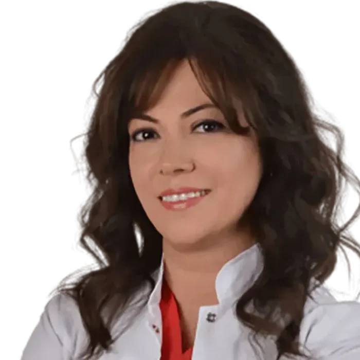
Dr Oksana Oturgan was born in Russia in 1987. In 2009 she graduated first in her class from the Ural State Medical Academy Faculty of Dentistry.
After deciding to continue her career in Turkey, she chose Marmara University for her specialisation and completed her endodontics residency there between 2015 and 2019. She then completed a PhD in endodontics at Istanbul Medipol University Faculty of Dentistry between 2021 and 2026, writing her thesis on the measurement of apical sealing in teeth with wide apices under the supervision of Prof. Mahir Günday. She regards having trained in the dental traditions of two different countries as an advantage that broadens her professional outlook.
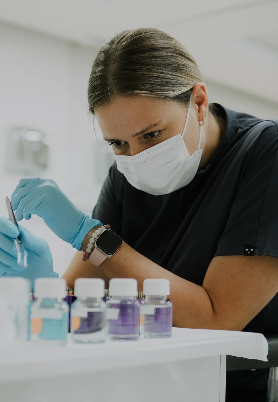
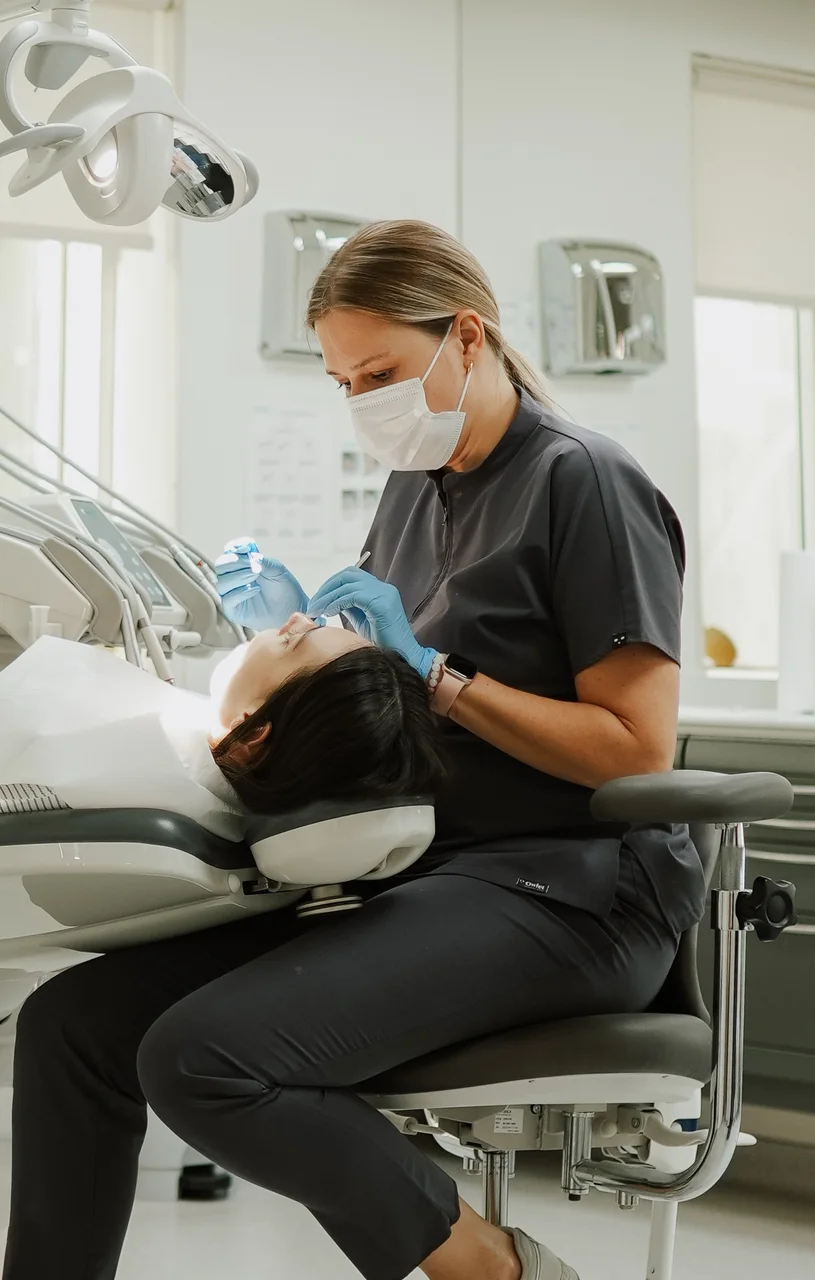
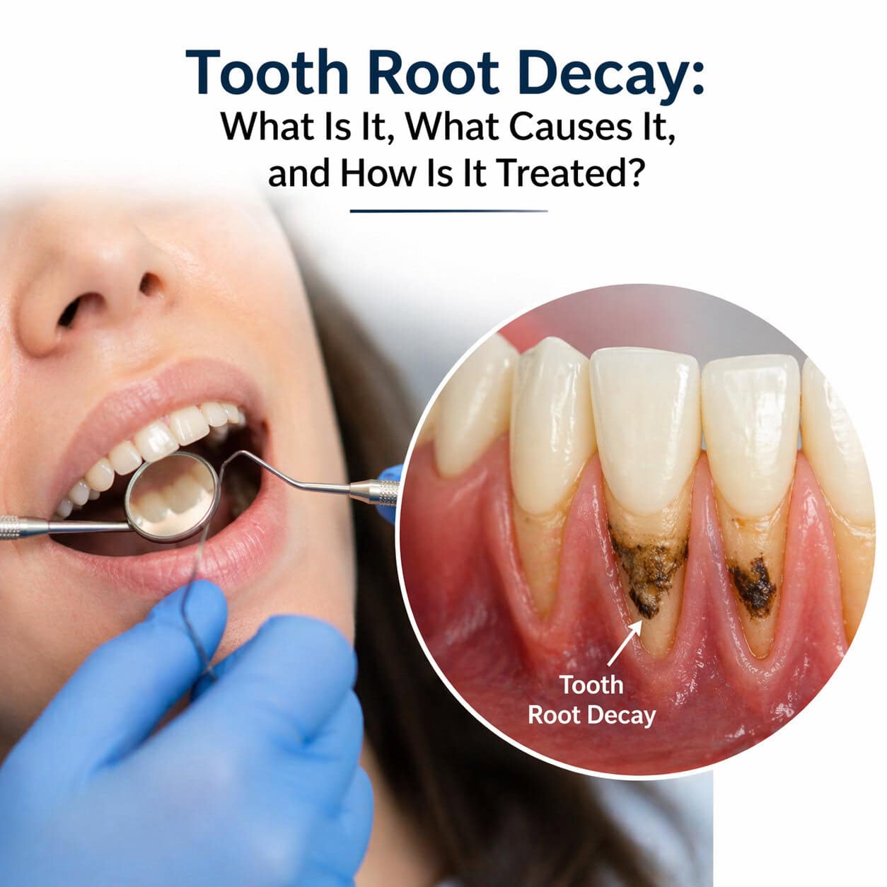
Micro-endodontics sits at the centre of her clinical work: she focuses on microscope-assisted root canal treatment, the renewal of previous root canal treatments (retreatment), MTA applications, vital pulp therapy and regenerative endodontics. She considers working under magnification indispensable, since the details the naked eye misses often decide the fate of a treatment.
She regularly attends advanced training programmes in endodontics and restorative dentistry, and closely follows developments in the techniques and materials of her field. She is a member of the Turkish Endodontic Society and the European Society of Endodontology (ESE).
Russian is her native language; she also speaks Turkish and English. Welcoming a significant share of patients from abroad in their own language, and running the whole treatment process in it, is a natural part of her international practice.
Outside dentistry she enjoys reading, trekking, yoga and pilates; she is interested in psychology and personal development, and travels both in Turkey and abroad.