
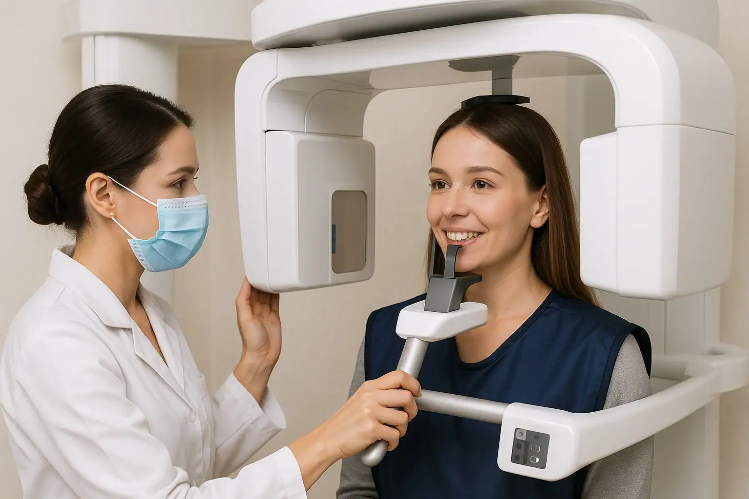
A panoramic dental X-ray, also called a panoramic radiograph or orthopantomogram, is a two-dimensional image that captures both jaws, all the teeth, the temporomandibular joints, the nasal area and the sinuses in a single rotating pass lasting roughly 10 to 20 seconds. It is the standard first look before wisdom tooth removal, implant placement and orthodontic planning, and for cysts, fractures and other jaw pathology. Doses are low: thermoluminescent dosimeter measurements in a phantom gave an effective dose of 17 microsieverts for a conventional panoramic unit and 8 microsieverts at the lowest digital exposure settings (PubMed 18448296).
- 01One rotating pass of about 10 to 20 seconds records both jaws, all the teeth, the temporomandibular joints, the nasal region and the sinuses in a single image.
- 02Measured effective dose in a Rando phantom was 17 microsieverts for a conventional panoramic device and 23 microsieverts for a digital one, falling to 8 microsieverts at the lowest exposure settings (PubMed 18448296).
- 03The panoramic view is a survey, not a detail image: small cavities and micro-cracks need intraoral bitewing or periapical films instead.
- 04Its commonest single indication is impacted teeth, particularly third molars, where root position drives the extraction plan.
- 05Preparation is limited to removing jewellery, glasses and metal objects; the patient bites a sterile guide and stays still while the arm rotates.
/ Introduction to Panoramic Dental X-Rays
Modern dentistry relies heavily on advanced imaging technologies to diagnose and treat oral health problems effectively. Among these technologies, the panoramic dental X-ray stands out as one of the most comprehensive and widely used diagnostic tools.
Unlike traditional intraoral X-rays that focus on a few teeth at a time, a panoramic x-ray captures the entire mouth including the upper and lower jaws, teeth, temporomandibular joints (TMJ), nasal area, and sinuses in a single image.
What Is a Panoramic Dental X-Ray?
A panoramic dental x-ray, also known as a panoramic radiograph or orthopantomogram (OPG), is a two-dimensional imaging technique that provides a broad view of the entire mouth in one shot.
This type of X-ray shows all teeth in both the upper and lower jaws, as well as their surrounding bones and supporting structures.
Because of its wide coverage, it’s often used for:
- Detecting impacted teeth or wisdom teeth
- Identifying jawbone abnormalities or cysts
- Evaluating sinus problems
- Planning orthodontic or implant treatments
- Assessing trauma, infections, or fractures
The digital panoramic x-ray version uses advanced sensors instead of traditional films, providing instant, high-resolution images with minimal radiation exposure.
/ How Panoramic X-Rays Work and Why They Are Needed
The panoramic x-ray machine consists of a rotating arm that moves around the patient’s head. This arm houses both an X-ray source and a sensor (or film) on opposite sides.
When activated, the arm rotates in a semicircular motion, capturing multiple images that are digitally combined into a single panoramic view.
Step-by-step procedure:
- Preparation
The patient is asked to remove jewelry, glasses, and metal objects that might interfere with the image.
- Positioning
The patient stands or sits in the X-ray unit, biting gently on a sterile plastic guide to keep the jaws steady.
- Exposure
The machine rotates around the head for about 10–20 seconds. During this time, the patient must stay completely still.
- Image generation
The digital panoramic x-ray system immediately processes and displays the image on the computer screen for the dentist’s evaluation.
This method is non-invasive, needs no anesthesia .
Why Is a Panoramic X-Ray Needed?
Dentists recommend a panoramic x-ray for several reasons, depending on each patient’s condition.
1. Comprehensive Oral Assessment
The panoramic view provides a complete picture of both jaws, all teeth, and surrounding tissues. It helps dentists detect underlying problems not visible during a routine clinical exam.
2. Wisdom Teeth and Impacted Tooth Evaluation
It’s one of the most common reasons for taking a panoramic dental x-ray. Impacted third molars (wisdom teeth) are clearly visible, helping dentists plan extraction or monitoring.
3. Orthodontic and Implant Planning
For orthodontic treatments or dental implants, panoramic radiographs help assess bone structure, tooth alignment, and root position, critical for precise planning.
4. Detection of Pathologies
A panoramic radiograph can reveal cysts, tumors, bone irregularities, infections, or sinus conditions that may require further attention.
5. Jaw Joint (TMJ) Assessment
The panoramic image shows the temporomandibular joints, helping in diagnosing TMJ disorders such as misalignment, arthritis, or jawbone erosion.
/ Advantages and Safety of Panoramic Dental X-Rays
1. Complete Coverage
Unlike intraoral X-rays that capture small sections, a panoramic X-ray shows the entire mouth, which is essential for accurate diagnosis and treatment planning.
2. Minimal Radiation Exposure
Modern digital panoramic x-rays emit far less radiation compared to traditional methods, making them safe even for children and frequent dental patients.
3. Quick and Non-Invasive
The process takes less than a minute, requires no discomfort, and provides immediate results.
4. Early Detection of Problems
Hidden infections, cysts, or bone loss can be identified before they cause pain or visible damage.
5. Essential for Complex Treatments
Whether planning an implant, evaluating jaw fractures, or preparing for orthodontic alignment, panoramic imaging provides a solid diagnostic foundation.
Are Panoramic X-Rays Safe?
Yes, panoramic x-rays are safe when performed by trained professionals using modern equipment.
The radiation dose is very low typically less than a day’s exposure to natural background radiation.
Safety measures include:
- Use of digital sensors that reduce radiation.
- Correct positioning, so the image does not have to be retaken.
- Strict adherence to ALARA (“As Low As Reasonably Achievable”) safety principles.
Lead aprons and thyroid collars are not on this list. The American Academy of Oral and Maxillofacial Radiology recommends discontinuing gonadal and fetal shielding, because the dose these areas receive from dental imaging is negligible, and recommends that thyroid shielding not be used for intraoral, panoramic, cephalometric or cone-beam CT imaging (PubMed 37530694). The American Dental Association notes that such shields can block the primary beam and lead to retakes (ada.org).
Lead aprons and thyroid collars are not on this list. The American Academy of Oral and Maxillofacial Radiology recommends discontinuing gonadal and fetal shielding, because the dose these areas receive from dental imaging is negligible, and recommends that thyroid shielding not be used for intraoral, panoramic, cephalometric or cone-beam CT imaging (PubMed 37530694). The American Dental Association notes that such shields can block the primary beam and lead to retakes (ada.org).
However, pregnant women should inform their dentist before any X-ray, as extra precautions or postponement might be necessary.
/ Panoramic Dental X-Ray Procedure and Limitations
The panoramic X-ray procedure is part of modern dental imaging and has practical limitations.
- No special preparation is required simply remove metallic accessories.
- The dentist or technician positions your head using chin and temple supports.
- You bite on a sterile guide to align teeth properly.
- The machine rotates around your head for about 15 seconds; you feel nothing and it stays quiet.
- The image appears instantly on the screen for analysis.
Afterward, your dentist will review the image to check for:
- Decay between teeth
- Bone loss
- Jaw misalignment
- Sinus issues
- Cysts or other pathologies
Limitations of Panoramic X-Rays
While a panoramic X-ray offers excellent overall views, it may not show fine details like small cavities or micro-cracks.
For those, intraoral bitewing X-rays may be needed.
Thus, panoramic imaging is often used as a first step in a comprehensive dental evaluation.
The Role of Panoramic X-Rays in Modern Dentistry
The use of digital panoramic X-rays has become a cornerstone in preventive and restorative dentistry.
From diagnosing hidden decay to planning complex prosthetic or implant procedures, it enhances both accuracy and patient safety.
Moreover, the ability to instantly view and share digital images helps dental teams collaborate efficiently and maintain accurate patient records.
/ Frequently Asked Questions (FAQ)
What is a panoramic dental x-ray used for?
It’s used to evaluate teeth, jaws, sinuses, and surrounding bone structures, often for diagnosis, treatment planning, or detecting abnormalities.
How often should I have a panoramic x-ray?
Typically every 3–5 years, or as recommended by your dentist, depending on your oral health needs.
Does a panoramic x-ray hurt?
No. It is quick, non-invasive, and you feel nothing while the machine moves.
Is a panoramic x-ray safe for children?
Yes, the radiation is minimal and digital panoramic systems are designed to adjust exposure levels for children.
Can panoramic x-rays detect cavities?
They can detect large decay areas but not very small cavities, intraoral X-rays are better for detailed tooth-by-tooth imaging.
What’s the difference between a panoramic x-ray and a regular dental x-ray?
A regular dental X-ray (bitewing or periapical) focuses on specific teeth, while a panoramic x-ray shows the entire mouth and jaw in one image.
How much does a panoramic dental x-ray cost?
Depending on the clinic and country, costs range from $30 to $150, but it’s often included in comprehensive dental check-ups.
In Mosdent dental clinics, this service is offered free of charge as part of the treatment.
Conclusion
The panoramic dental x-ray is an essential diagnostic tool that provides a complete view of your oral and maxillofacial structures.
It’s safe, quick, and highly informative, allowing dentists to detect potential issues early and plan treatments accurately.
Whether you’re considering orthodontic care, dental implants, or a routine exam, a panoramic x-ray ensures that nothing is overlooked safeguarding both your oral health and overall well-being.
Let’s plan the right treatment together.
Free Assessment→- 1.Gavala S, Donta C, Tsiklakis K, Boziari A, Kamenopoulou V, Stamatakis HC. Radiation dose reduction in direct digital panoramic radiography. Eur J Radiol 2009;71:42-8. PubMed 18448296
- 2.Benavides E, Bhula A, Gohel A, Lurie AG, Mallya SM, Ramesh A, et al. Patient shielding during dentomaxillofacial radiography: Recommendations from the American Academy of Oral and Maxillofacial Radiology. J Am Dent Assoc 2023;154:826-835.e2. PubMed 37530694
- 3.American Dental Association. X-Rays/Radiographs. Oral Health Topics, ADA Library & Archives; topic last updated 26 March 2026 (accessed 11 September 2026)