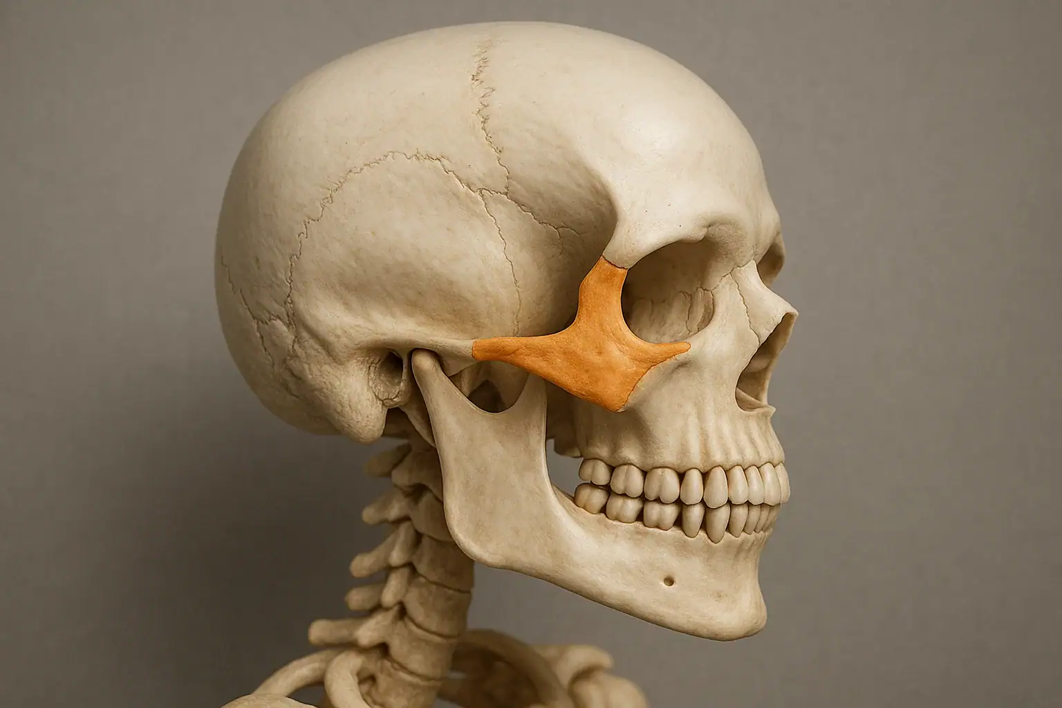
The zygomatic bone, also called the cheekbone, malar bone or zygoma, is a paired irregular facial bone that gives the cheek its shape and forms part of the lateral wall and floor of the eye socket. It articulates with four bones: the frontal bone, the maxilla, the temporal bone and the sphenoid. Its temporal process meets the zygomatic process of the temporal bone to form the zygomatic arch, the bridge the masseter attaches to and the temporalis passes beneath. Ossification begins around the eighth week of fetal development. Because the bone sits forward and to the side, it is a frequent site of facial fracture.
- 01The zygomatic bone forms joints with four bones, the frontal, maxilla, temporal and sphenoid, which is why a displaced fracture shows up as flattened cheek, double vision and difficulty opening the mouth at the same time.
- 02The zygomatic arch is not a separate bone: it is the temporal process of the zygoma joined to the zygomatic process of the temporal bone, and it carries the masseter while the temporalis runs underneath it.
- 03Zygomaticomaxillary complex fracture is the clinical name for the common injury pattern. In a series of 468 patients, 400 were male and 68 female, mean age 37.1 years, and assaults accounted for 64.5 per cent of cases (PubMed 20677370).
- 04Treatment follows displacement: minor undisplaced fractures can be managed without surgery, displaced ones are plated. In that same 468-patient series 96.6 per cent were treated surgically, most often with miniplate osteosynthesis (PubMed 20677370).
- 05The bone anchors zygomaticus major and minor, the muscles that lift the corner of the mouth and the upper lip, so its position affects how a smile moves as well as how the midface looks.
The human skull is a structural masterpiece, and among its most recognizable features is the zygomatic bone, also commonly known as the cheekbone. Not only does it define the contours of the midface, but it also supports essential functions including protection of the eye socket, attachment for facial muscles, and articulation with surrounding cranial structures.
This guide covers what you need to know about the zygomatic bone: its anatomy, articulations, development, clinical relevance, and aesthetic importance. Whether you're studying anatomy, pursuing dental or medical education, or simply curious about facial bones, this article provides the complete picture.
/ Zygomatic Bone Anatomy and Location
What Is the Zygomatic Bone?
The zygomatic bone is a paired, irregular facial bone that gives shape to the cheeks and forms part of the lateral wall and floor of the orbit (eye socket). It is often referred to as:
- Cheekbone
- Malar bone
- Zygoma
It plays a vital role in both form and function by contributing to facial aesthetics and serving as a structural foundation for surrounding tissues.
Where Is the Zygomatic Bone Located?
The zygomatic bones are situated on each side of the face and are easily palpable just below and to the side of the eyes.
Anatomical Position:
- Inferior to the orbit
- Lateral to the nasal bones
- Anterior to the temporal bones
- Superior to the maxillae
These bones give the cheeks their prominence and serve as lateral buttresses for the midface.
Zygomatic Bone Anatomy: Detailed Structure
Each zygomatic bone has several projections (or processes) that articulate with neighboring bones, contributing to the structural complexity of the skull.
1. Temporal Process of the Zygomatic Bone
- Projects posteriorly to connect with the zygomatic process of the temporal bone .
- This connection forms the zygomatic arch, a crucial anatomical feature of the skull.
2. Zygomatic Process of the Temporal Bone
- A bony projection of the temporal bone that completes the zygomatic arch.
- Provides a path for the temporalis muscle, essential for mastication.
3. Frontal Process
- Extends superiorly to articulate with the frontal bone near the lateral aspect of the orbit.
4. Maxillary Process
- Projects medially and joins the maxilla, contributing to the infraorbital rim and orbital floor.
Articulations of the Zygomatic Bone
The zygomatic bone forms joints with four bones:
| Bone | Connected Process |
|---|---|
| Frontal bone | Frontal process of zygomatic bone |
| Maxilla | Maxillary process |
| Temporal bone | Temporal process & zygomatic process |
| Sphenoid bone | Orbital surface |
These articulations support the orbit, cheek, and jaw structures and create stability across the midface.
/ Zygomatic Bone Function and Muscle Attachments
Key Functions of the Zygomatic Bone
The zygomatic bones are essential for both aesthetic and functional reasons:
- ✅ Facial contour and projection
- ✅ Orbit protection
- ✅ Attachment site for facial muscles like the zygomaticus major and minor
- ✅ Chewing efficiency via the zygomatic arch’s relation to the masseter and temporalis muscles
- ✅ Trauma absorption due to their lateral, prominent position
Muscles Attached to the Zygomatic Bone
Several muscles responsible for facial expression and mastication originate or insert on the zygomatic bone:
- Zygomaticus major – Lifts the corner of the mouth (smiling)
- Zygomaticus minor – Elevates the upper lip
- Masseter – Attaches to the zygomatic arch, aids in chewing
- Temporalis – Passes beneath the zygomatic arch, controls jaw closure
Zygomatic Arch: Structure and Role
The zygomatic arch is formed by the union of:
- Temporal process of the zygomatic bone
- Zygomatic process of the temporal bone
It serves to:
- Support chewing muscles
- Create facial width and definition
- Act as a landmark in reconstructive surgery and trauma cases
Development and Ossification
- The zygomatic bone begins ossification during the 8th week of fetal development from neural crest cells.
- It is completely ossified in early childhood and fuses with neighboring bones to form the mature midface.
/ Zygomatic Bone Clinical Significance
Zygomatic Bone in Cosmetic and Reconstructive Procedures
1. Cheek Augmentation
- Enhances midface projection using:
- Solid implants (silicone or Medpor)
- Hyaluronic acid fillers
- Fat grafting
2. Zygoma Reduction
- Common in Asian facial contouring surgeries to slim the face by reducing lateral cheek projection.
3. Zygomatic Implants
- Used in patients with severe maxillary bone loss where traditional dental implants are not feasible.
Clinical Conditions and Fractures
Zygomaticomaxillary Complex (ZMC) Fracture
A common facial injury involving:
- Zygomatic bone
- Maxilla
- Orbit
- Lateral orbital rim
Symptoms include:
- Flattened cheek
- Facial asymmetry
- Diplopia (double vision)
- Paresthesia due to infraorbital nerve compression
- Difficulty opening the mouth
Treatment Options:
- Non-surgical : For minor, non-displaced fractures
- Surgical : Open reduction and internal fixation (ORIF) with titanium plates and screws
Plating a displaced cheekbone is maxillofacial work, not general dentistry. At Mosdent it is handled under treatment of jaw fracture, alongside the rest of our oral surgery service.
Imaging and Diagnostic Tools
- X-ray – Basic evaluation
- CT Scan – Preferred for trauma and surgical planning
- CBCT (Cone Beam CT) – High-resolution imaging in dental implantology
- MRI – For adjacent soft tissue involvement or tumor evaluation
Anthropological and Forensic Relevance
- Zygomatic bones are used to determine ancestry, biological sex, and age of skeletal remains.
- Prominent cheekbones are often associated with East Asian and Native American ancestry.
- Facial reconstruction uses the zygoma as a key anchor point.
Variations Across Populations
Different ethnic populations display unique zygomatic morphologies, which can impact both facial aesthetics and surgical planning.
| Population | Zygomatic Characteristics |
|---|---|
| East Asian | Highly prominent, laterally flared |
| European | Balanced projection, moderate prominence |
| African | Broad, robust, and anteriorly angled |
/ Frequently Asked Questions (FAQs)
What is the zygomatic bone?
It is a paired facial bone also known as the cheekbone, forming the prominence of the cheek and part of the eye socket.
What is the temporal process of the zygomatic bone?
A projection that connects with the zygomatic process of the temporal bone to form the zygomatic arch.
Where is the zygomatic bone located?
On the upper lateral side of the face, between the maxilla and the temporal bone, beneath the eye.
What is the zygomatic process of the temporal bone?
A projection of the temporal bone that articulates with the zygomatic bone.
Can a fractured zygomatic bone heal without surgery?
Mild, non-displaced fractures may heal on their own, but many require surgical intervention for function and facial symmetry.
How is the zygomatic bone involved in dental implantology?
Zygomatic implants are anchored into the bone when maxillary bone mass is insufficient for traditional implants.
/ Summary Table and Conclusion
Summary Table
| Aspect | Details |
|---|---|
| Common Names | Zygoma, cheekbone, malar bone |
| Location | Lateral midface, below the eye socket |
| Processes Involved | Temporal, maxillary, frontal, orbital |
| Articulated Bones | Maxilla, temporal, sphenoid, frontal |
| Related Muscles | Zygomaticus major/minor, masseter, temporalis |
| Clinical Relevance | Fractures, implants, cosmetic surgery, facial reconstruction |
| Diagnostic Tools | CT, CBCT, X-ray, MRI |
| Development Origin | Neural crest, ossifies during fetal development |
| Anthropological Significance | Used in forensic identification and craniofacial classification |
Conclusion
The zygomatic bone is more than just the prominent curve of your cheek, it’s a crucial architectural, functional, and aesthetic cornerstone of the human face. It interacts with multiple bones and muscles, affects dental and ophthalmologic outcomes, and holds a prominent role in cosmetic, reconstructive, and forensic practices.
Published trauma series describe who sustains cheekbone fractures and how they are repaired. In a Polish series of 468 patients treated for zygomaticomaxillary complex fractures, 400 were men and assault caused 64.5% of the injuries. Zingg type B fractures made up 73.1% of cases, and miniplate osteosynthesis was the most favored technique (PubMed 20677370). A prospective CBCT study followed 50 adults treated with open reduction and internal fixation. Fixation at 4 points gave more facial symmetry than 2 or 3 points, and chronic sinusitis after surgery was uncommon (PubMed 37336318).
Whether you're a healthcare professional, anatomy student, or cosmetic surgeon, understanding the zygomatic bone, including its temporal process, zygomatic arch, and clinical implications, provides essential insight into facial harmony, structure, and health.
Let’s plan the right treatment together.
Free Assessment→- 1.Bogusiak K, Arkuszewski P. Characteristics and epidemiology of zygomaticomaxillary complex fractures. J Craniofac Surg 2010;21:1018-23. PubMed 20677370
- 2.Al-Shujaa EA, Al-Awdi HH, Al-Wesabi SN, Lubamba GP, Altayar BA, Sakran KA, et al. Maxillary sinus volume and facial asymmetry after unilateral zygomaticomaxillary complex fracture reduction with 2, 3, and 4 fixation points: CBCT-based investigation. J Stomatol Oral Maxillofac Surg 2024;125:101537. PubMed 37336318