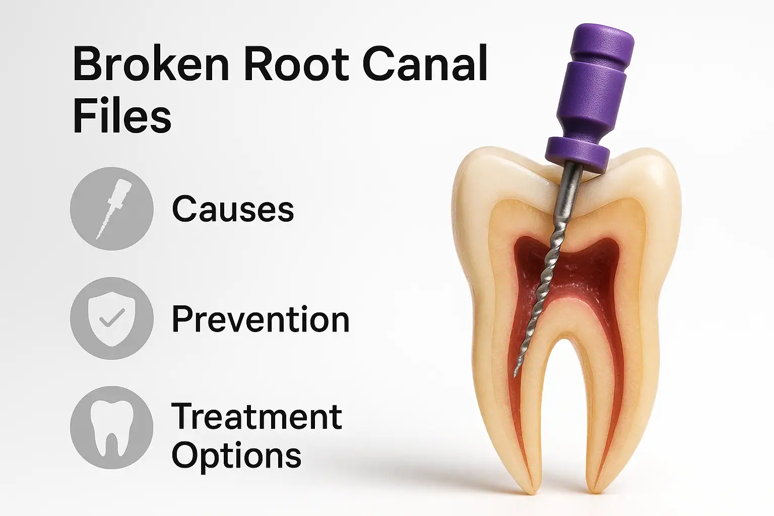
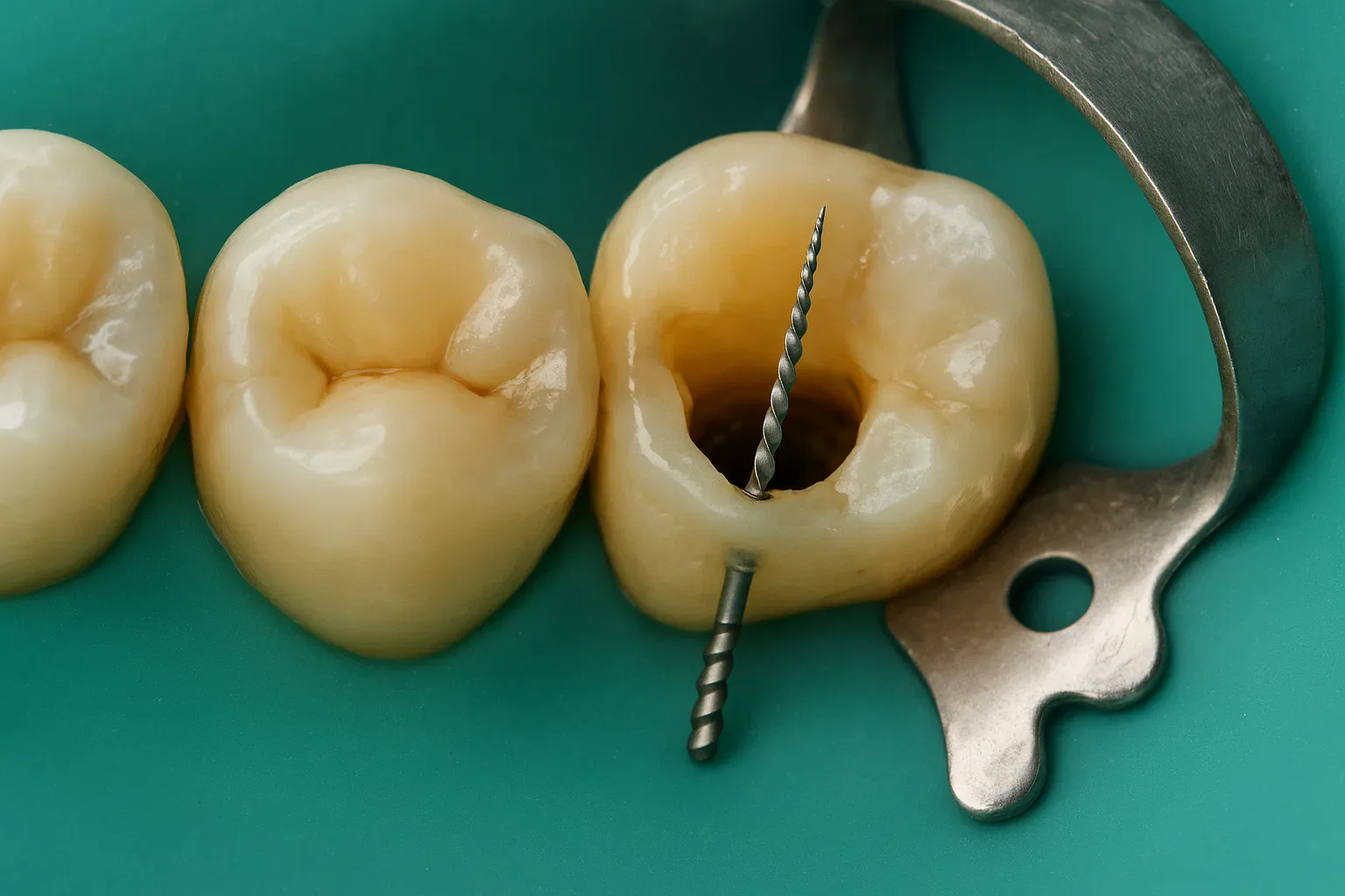
A broken endodontic file, clinically a separated instrument, is a fragment of a root canal file that fractures during shaping and stays inside the canal, most often in the apical third or in a curved segment. It is a recognised procedural incident rather than a rare accident: a systematic review with meta-regression of clinical studies pooled the fracture incidence of engine-driven nickel-titanium files at 2.27 percent, 2.43 percent with continuous rotation and 1.0 percent with reciprocation (PubMed 33410578). A retained fragment does not by itself condemn the tooth; management depends on the fragment's position and length and on the state of the canal.
- 01Separated instrument means a file fragment left inside the canal; pooled clinical incidence for engine-driven nickel-titanium files was 2.27 percent, with 2.43 percent for rotary and 1.0 percent for reciprocating motion (PubMed 33410578).
- 02Prognosis is driven by the infection rather than by the metal: across 199 matched cases, weighted mean healing with a retained fragment was 91 percent and the risk difference against conventional cases was 0.01 (PubMed 20416418).
- 03The pre-existing periapical lesion is the stronger predictor: 80.7 percent of teeth with a lesion healed, compared with 92.4 percent of teeth that started without one (PubMed 20416418).
- 04Meta-regression identified two operator-side factors linked to higher fracture incidence: using the same nickel-titanium files in more than four teeth, and lower operator proficiency (PubMed 33410578).
- 05Three routes exist once a file separates: ultrasonic retrieval under the microscope, bypassing the fragment to clean and fill the rest of the canal, or leaving it in place under radiographic follow-up, with apical surgery as the fallback.
Endodontic (root canal) treatment is one of the most common procedures in dentistry. However, during this procedure, the instruments used to clean and shape the root canal system, called files, may fracture inside the canal. This “broken endodontic file” situation can be challenging for both the dentist and the patient.
In this comprehensive guide, we will examine why file separation occurs, its risk factors, prevention methods, and treatment options.
/ Broken Endodontic Files and Their Causes
What Is a Broken Endodontic File?
A root canal file is a metal instrument used to shape and clean the inside of the root canal. These files are typically made of stainless steel or nickel-titanium (NiTi) alloy.
A broken file refers to the situation where a file fractures during treatment, leaving a portion of it inside the root canal. This fragment may block the canal and prevent complete cleaning or filling.
In many cases, file separation occurs in the apical third of the canal (near the root tip) or in curved areas, which are anatomically narrower and harder to access, making removal more complex.
Causes of Broken Endodontic Files
1. Excessive Mechanical Stress
- Root canal files are very thin and flexible, but when too much force is applied, the risk of distortion or fracture increases.
- The risk is even higher in narrow, curved canals.
- Inadequate preparation of a glide path increases the stress on the file.
2. Metal Fatigue
- NiTi instruments are flexible but are prone to cyclic fatigue when used repeatedly.
- Overusing files beyond their recommended life cycle significantly raises the risk of breakage.
- Repeated autoclaving and heat exposure can further weaken the metal structure.
3. Improper Speed and Torque Settings
- Ignoring the manufacturer’s recommended rotational speed and torque settings may compromise file integrity.
- Excessive speed or low torque can overload the instrument, leading to fracture.
4. Insufficient Irrigation and Lubrication
- If the canal is not adequately irrigated, friction increases, which promotes file separation.
- Proper lubrication also helps flush debris away, reducing torsional stress on the file.
5. Tooth Anatomy
- Curved, narrow, calcified, or S-shaped canals significantly increase the risk of instrument fracture.
- In elderly patients, canals tend to be more calcified and constricted, raising the difficulty level.
/ File Separation Risks and Diagnosis
Risk Factors for File Separation
- Reusing files multiple times despite visible wear
- Operating rotary instruments at excessively high speed for prolonged periods
- Inexperience or inadequate training of the operator
- Insufficient radiographic evaluation and lack of 3D planning
- Applying excessive apical pressure
Additionally, improper working length determination can cause the file to bind apically, increasing fracture risk.
Symptoms and Diagnosis
Most of the time, the dentist notices the instrument separation during the procedure.
However, some patients may present later with pain, swelling, or persistent discomfort .
Diagnostic Tools:
- Periapical radiographs are used first.
- CBCT (Cone Beam Computed Tomography) may be taken to precisely locate the broken file fragment.
- Visual inspection under a dental operating microscope can confirm the position and orientation.
Accurate diagnosis is crucial to plan the next steps and select the most suitable treatment approach.
Potential Consequences of a Broken File
A separated file can compromise the success of root canal treatment.
Possible Outcomes:
- Incomplete cleaning and shaping of the canal
- Persistent bacterial infection
- Failure of periapical lesion healing
- Increased risk of tooth loss
However, in many cases, the tooth can still be treated successfully and remain functional for many years, especially if the issue is managed promptly.
/ Prevention and Treatment Options for a Broken File
Prevention Methods
1. Regular Instrument Inspection
Files should be carefully examined visually and under magnification before each use. Any distortion or unwinding should be a reason for disposal.
2. Follow Manufacturer’s Guidelines
- Adhere strictly to recommended speed and torque values .
- Avoid reusing single-use files.
3. Adequate Irrigation
Maintain continuous irrigation with sodium hypochlorite, EDTA, or other recommended solutions to keep the canal moist and reduce friction.
4. Glide Path Creation
Establish a smooth, reproducible glide path with hand files before using rotary instruments. This reduces torsional stress and lowers fracture risk.
5. Proper Training and Experience
Experienced clinicians are better able to anticipate challenging canals and select the correct file sequence accordingly.
Additional precautions include radiographic planning , use of crown-down or step-back techniques , and avoiding excessive apical pressure during preparation.
Treatment Options for a Broken File
The management of a separated instrument depends on its location, the length of the fragment, and the condition of the canal.
1. File Retrieval
Whenever possible, the fragment is removed.
- Ultrasonic tips under microscopic vision are used to vibrate and loosen the fragment.
- Specialized retrieval kits and Hedström files may be employed.
- Once removed, the canal is re-instrumented and disinfected thoroughly.
Retrieval under magnification is a different discipline from routine root canal work. At Mosdent it runs as micro endodontic retreatment, with the operating microscope and ultrasonic tips as standard rather than optional equipment.
2. Bypass Technique
If removal is not feasible, the canal may be negotiated around the fragment, allowing cleaning and filling of the remainder of the canal.
3. Obturation and Monitoring
In some cases, the file is left in place and the canal is filled as best as possible. The tooth is then monitored radiographically over time.
4. Surgical Treatment
If the fragment is located in the apical region and cannot be removed non-surgically, apicoectomy and retrograde filling may be performed.
/ Managing File Separation Successfully
Importance of Microscope and CBCT
Modern dentistry relies heavily on dental operating microscopes and CBCT imaging for successful management of separated instruments.
Benefits:
- Precise visualization of the fragment’s position
- Minimally invasive removal techniques
- Preservation of surrounding dentin structure
- Higher success rates with fewer complications
Patient Responsibilities
- Attending follow-up appointments after treatment
- Maintaining good oral hygiene to prevent reinfection
- Reporting pain, swelling, or any unusual symptoms promptly
- Keeping radiographs or CBCT scans for reference if consulting another specialist
Success Rate of Treatment
Success depends on multiple factors, including the fragment’s location, operator experience, and available technology.
The use of operating microscopes and ultrasonics has significantly improved success rates.
Prompt intervention and careful planning increase the chances of saving the tooth.
Conclusion
Broken endodontic files are one of the most complex complications in root canal treatment, but thanks to modern techniques, they are no longer a hopeless scenario.
- Early detection and careful treatment planning significantly improve prognosis.
- The use of operating microscopes , CBCT imaging , and ultrasonic systems allows for safer and more predictable removal or bypassing of separated files.
- Collaboration between dentist and patient, through compliance with treatment recommendations and regular follow-ups, plays a key role in long-term success.
Encountering a broken file does not necessarily mean losing the tooth. With the right approach and technology, the tooth can often be preserved for many years, maintaining function and aesthetics.
/ FAQs
Does a broken file mean I will lose my tooth?
No. In many cases, the file can be retrieved or bypassed, allowing the tooth to be treated and kept functional.
Can a broken file cause pain?
It may cause pain if it leads to persistent infection. Timely follow-up is essential.
How much does broken file treatment cost?
The use of a dental microscope and advanced equipment may increase the cost, but it is often worth it to save the tooth.
What happens if the file is not removed?
Sometimes the fragment is left in place and the tooth is monitored. If infection develops later, surgical intervention may be required.
How long does it take to remove a broken file?
Depending on the case complexity, it may take a single appointment or several visits. Microscope assistance usually shortens the procedure time.
Let’s plan the right treatment together.
Free Assessment→- 1.Panitvisai P, Parunnit P, Sathorn C, Messer HH. Impact of a retained instrument on treatment outcome: a systematic review and meta-analysis. J Endod 2010;36:775-80. PubMed 20416418
- 2.Gomes MS, Vieira RM, Böttcher DE, Plotino G, Celeste RK, Rossi-Fedele G. Clinical fracture incidence of rotary and reciprocating NiTi files: A systematic review and meta-regression. Aust Endod J 2021;47:372-385. PubMed 33410578