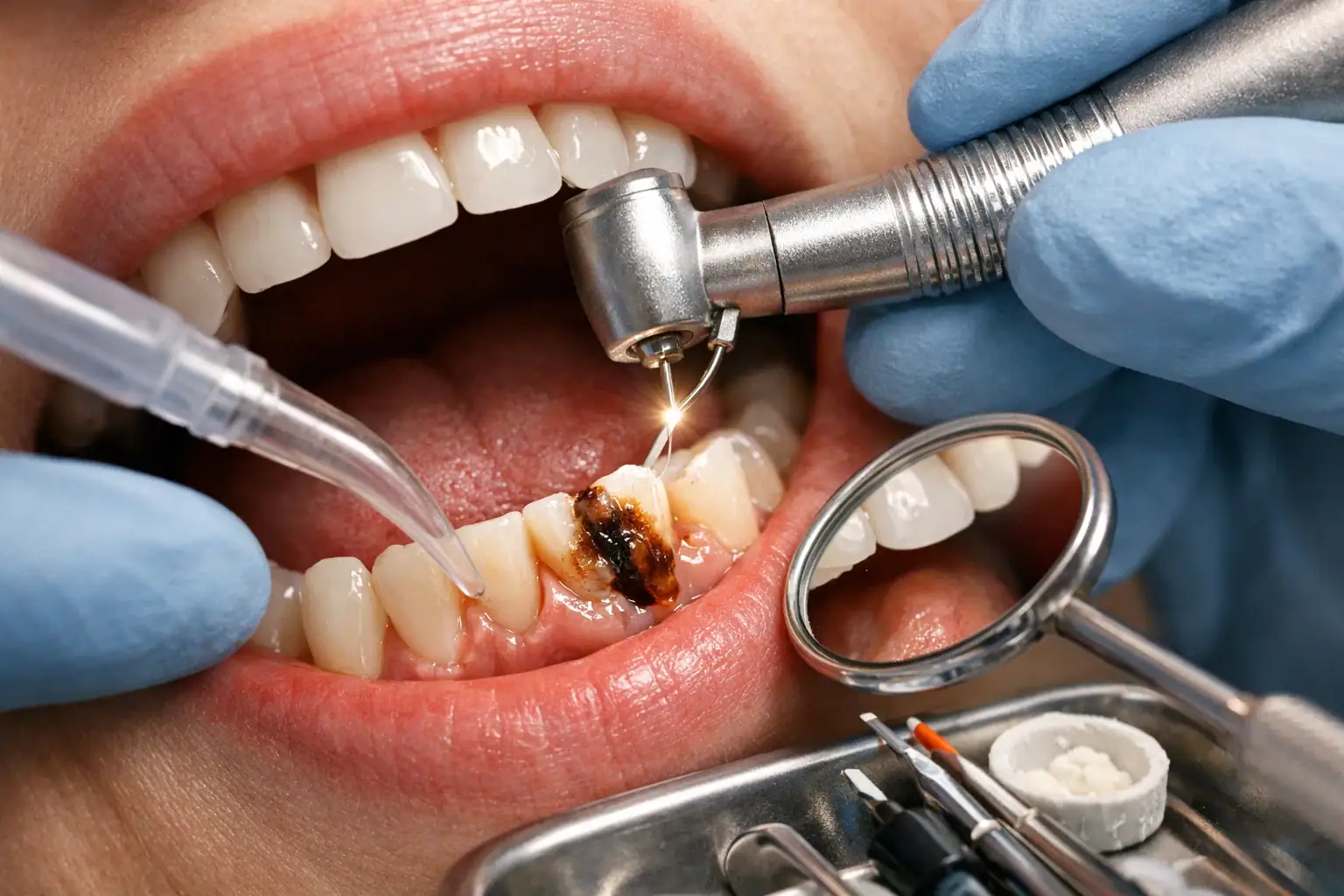
A tooth root cavity, or root caries, is decay on the cementum and dentine of the root surface, which is exposed once the gum recedes and has no enamel layer to protect it. Across 74 published surveys, pooled prevalence is 41.5% of adults (PubMed 30705564). Early lesions may be arrested with fluoride varnish, high-fluoride toothpaste or silver diamine fluoride; once the surface is cavitated, the decay is cleaned out and restored, often with a fluoride-releasing glass ionomer. Lesions reaching the pulp need root canal treatment, and unrestorable teeth are extracted.
- 01Root caries is decay of cementum and dentine at or below the gum margin; unlike a crown cavity there is no enamel layer above it to slow the acid attack.
- 02Pooled prevalence is 41.5% of adults across 74 surveys (PubMed 30705564); a separate 91-publication analysis of middle-aged and elderly groups reports 41% prevalence and a mean of 2.87 affected teeth (PubMed 39681109).
- 03Silver diamine fluoride probably prevents new root caries, with a mean difference of -0.79 surfaces against placebo or no treatment (95% CI -1.40 to -0.17; 3 studies, 439 participants, moderate-certainty evidence; PubMed 39508296). Its known drawback is black staining of the arrested lesion.
- 04Glass ionomer is often chosen for root cavity fillings because it bonds chemically to tooth structure, tolerates the moisture found at the gum margin and releases fluoride over time.
- 05Root canal treatment enters the picture only when decay reaches the pulp; earlier stages are handled with fluoride therapy, silver diamine fluoride or a restoration.
/ Introduction: Why Root Cavities Deserve Special Attention
Most cavities form on the chewing surfaces or between teeth, areas covered by enamel. But root cavities are different. They develop on the root surface of the tooth, usually near or below the gumline, and they can progress faster than “regular” cavities because the root is not protected by enamel.
A tooth root cavity (often called root caries or root decay) is especially common in adults with gum recession, dry mouth, or a history of gum disease . These risk factors are becoming more widespread as people keep their natural teeth longer into older age.
This guide explains:
- what a tooth root cavity is (and what it looks like),
- what causes root cavities,
- how dentists diagnose root decay,
- the full range of root caries treatment options (including SDF and restorative approaches),
- and how to prevent root cavities from coming back.
What Is the Tooth Root, and Why Is It Easier to Decay?
A tooth has two main parts:
- Crown: the visible part above the gumline, protected by enamel (the hardest substance in the body).
- Root: the part anchored in the bone, covered by cementum and underlying dentin.
Unlike enamel, cementum and dentin are less mineralized and are more vulnerable to acid attack. Root surfaces can start demineralizing at higher pH levels than enamel, making them more prone to decay when exposed.
Key takeaway: When the gumline recedes and root surfaces become exposed, the tooth becomes much more susceptible to root decay.
/ Tooth Root Cavity Overview
What Is a Tooth Root Cavity (Root Caries)?
A tooth root cavity is a decayed area on the root surface of a tooth. It typically forms:
- along the gumline, or
- just under the gum margin, where plaque can accumulate and cleaning is harder.
Root cavities often:
- spread wider along the root surface,
- progress faster than enamel cavities,
- and can compromise the tooth’s structure quickly if not treated early.
Because root caries may be hidden under the gumline, some people don’t notice a problem until sensitivity or pain develops.
Root Cavities vs. Regular Cavities: The Practical Differences
Root cavities (root caries) differ from crown cavities in clinically important ways:
- Less protection : No enamel on the root.
- Faster progression : Softer tissues demineralize more easily.
- Harder restoration : Moisture control near the gumline is challenging; bonding can be more difficult.
- Higher recurrence risk : Gum recession, dry mouth, and plaque retention can persist.
What Does a Root Cavity Look Like?
A tooth root cavity may appear as:
- a yellowish, brown, or black area near the gumline,
- a soft or “leathery” spot when probed by a dentist,
- a shallow “scooped out” lesion on the root surface,
- or a darkened line at the gum margin that keeps expanding.
Important: Not every dark spot is decay. Root surfaces can stain. That’s why dentists confirm with careful exam and X-rays when needed.
/ Causes of Root Cavities
Root cavities are a classic example of multifactorial disease, a mix of bacteria, diet, saliva, tooth anatomy, and patient habits. The ADA describes dental caries as a biofilm-mediated, sugar-driven, multifactorial, dynamic disease.
1) Gum Recession
Root cavities usually start when gum recession exposes the root surface. Gum recession may be caused by:
- periodontal (gum) disease,
- aggressive brushing,
- thin gum tissue,
- teeth grinding (bruxism),
- orthodontic movement,
- tobacco use,
- aging-related changes.
Gum recession and root caries are tightly linked in the aging population.
2) Dry Mouth (Xerostomia)
Saliva helps:
- neutralize acids,
- provide minerals for remineralization,
- and wash away food debris.
When saliva is reduced (medications, dehydration, systemic conditions), the risk of root decay rises sharply.
3) Plaque Retention Near the Gumline
Root surfaces are harder to clean. Plaque can build up:
- around gumline notches,
- near crowns/bridges,
- between teeth (interproximal areas),
- around partial dentures or orthodontic retainers.
4) Frequent Sugary or Acidic Snacking
Root caries is often driven by frequency (how often you snack), not just the amount of sugar. Frequent exposure fuels acid attacks repeatedly.
5) Existing Restorations and Dental Work
Margins of crowns, bridges, veneers, and older fillings can create plaque-retentive edges. This can increase risk of root cavities especially at crown margins near the gumline.
6) Age, Health Conditions, and Dexterity
Root caries is more common in older adults, partly because recession, dry mouth, and chronic conditions become more prevalent. Some studies note high prevalence in older age groups.
/ Root Caries Risk and Diagnosis
Who Is Most at Risk for Root Caries?
You may be at higher risk of tooth root cavities if you have:
- gum recession or periodontal history,
- dry mouth (xerostomia),
- diabetes or multiple chronic conditions (multimorbidity),
- a high-sugar diet or frequent snacking,
- limited dexterity or inconsistent brushing/flossing,
- exposed roots from over-brushing,
- smoking or vaping,
- existing crowns/bridges and difficult-to-clean margins.
(Recent research also explores the link between multimorbidity and root caries among older adults.)
How Dentists Diagnose Root Cavities
1) Visual and Tactile Exam
Dentists check:
- gum recession patterns,
- plaque accumulation areas,
- root surface texture (soft/leathery),
- lesion color and boundaries.
2) Gentle Air-Drying and Isolation
Drying helps reveal early lesions and boundary changes.
3) Dental X-rays
X-rays help detect:
- interproximal root cavities,
- subgingival extension,
- proximity to pulp (whether root canal risk is higher).
4) Risk Assessment
A good dentist doesn’t only treat the lesion, they treat the cause:
- diet frequency,
- saliva status,
- fluoride exposure,
- plaque control,
- and gum health .
Staging Root Cavities: Early vs. Advanced Root Decay
A simple practical staging approach:
Early (Non-cavitated / Incipient)
- surface changes, mild discoloration,
- no obvious “hole,”
- may be arrestable with fluoride/remineralization and behavior change.
Moderate (Cavitated but Restorable)
- a clear defect or softened area,
- typically needs a filling (restoration).
Advanced (Deep Root Cavity)
- extensive decay close to the pulp,
- may require root canal treatment and full coverage (crown).
Hopeless (Non-restorable)
- structural compromise, fracture risk,
- deep decay undermining the tooth,
- extraction may be necessary.
/ Root Cavities Treatment and SDF
Root Cavities Treatment: What Works and Why
Treatment depends on lesion depth, activity, and patient risk factors (recession, dry mouth, diet).
1) Non-Invasive Treatment for Early Root Cavities
If a tooth root cavity is detected early, dentists may aim to arrest or reverse the process.
Professional Fluoride Varnish
High-concentration fluoride varnish can help:
- remineralize root surfaces,
- reduce sensitivity,
- slow lesion progression,
- protect adjacent exposed roots.
Prescription High-Fluoride Toothpaste
In many high-risk patients, clinicians recommend 5,000 ppm fluoride toothpaste (where available/prescribed). This is especially useful for:
- dry mouth,
- multiple exposed roots,
- recurrent root caries.
Remineralization Support + Plaque Control
- improved brushing technique,
- interdental cleaning (floss/interdental brushes),
- targeted cleaning at gumline,
- diet frequency control.
Reality check: Only very early lesions may be arrested or partially reversed. Once cavitated, restorative treatment is typically needed.
2) Silver Diamine Fluoride (SDF) for Root Caries
Silver diamine fluoride (SDF) is a topical agent used to arrest caries, including root caries. It has both antimicrobial (silver) and remineralizing (fluoride) actions.
When SDF Is Especially Helpful
- Older adults with multiple root lesions
- Patients who can’t tolerate long treatment visits
- High-caries-risk individuals (dry mouth, limited hygiene ability)
- Situations where “stop the disease now” is the priority
Important Drawback: Staining
SDF commonly causes black staining of the arrested lesion. For posterior or non-esthetic areas, this may be acceptable. For front teeth, dentists may discuss alternatives or combined approaches.
Evidence Snapshot
Cochrane evidence suggests SDF probably helps prevent new root caries compared to placebo/no treatment (though evidence for some comparisons remains uncertain).
/ Root Caries Treatment Options
3) Fillings for Root Cavities (Restorative Treatment)
Once a root cavity is cavitated, the dentist typically removes decayed tissue and restores the area.
Why Root Cavity Fillings Are Tricky
- proximity to the gumline (bleeding control)
- moisture contamination (saliva, crevicular fluid)
- limited enamel for bonding
- margin placement sometimes below the gum
Best Materials for Root Caries Treatment
Glass Ionomer Cement (GIC)
Often preferred for root cavities because it:
- bonds chemically to tooth structure,
- tolerates moisture better than composite,
- releases fluoride over time,
- can reduce recurrent decay risk.
Resin-Modified Glass Ionomer (RMGIC)
Improved strength and handling while still offering fluoride release.
Composite Resin
Provides better esthetics and polish, but requires excellent isolation and bonding technique, sometimes difficult near the gum.
Clinical tip: In high-risk patients, fluoride-releasing materials are often advantageous for root cavity treatment.
4) When Does a Tooth with a Root Cavity Need a Crown?
A crown may be recommended if:
- decay is extensive and undermines tooth structure,
- a large portion of the tooth is restored,
- the tooth is cracked or structurally compromised,
- a root canal has been performed (often recommended for protection).
Sometimes a crown lengthening procedure ( periodontal surgery ) is needed to expose more tooth structure so a crown margin can be placed properly.
5) Root Canal Treatment for Root Decay
If root cavities reach the pulp (nerve), bacteria can inflame or infect the pulp leading to:
- lingering pain,
- spontaneous toothache,
- swelling,
- abscess risk.
In these cases, root canal treatment is used to:
- remove infected pulp,
- disinfect canals,
- seal the tooth,
- and then restore with a filling/crown.
6) Extraction and Replacement (Last Resort)
If root decay is too advanced to restore, extraction may be necessary. Replacement options include:
- dental implant,
- bridge,
- partial denture .
Root caries, when left untreated, can lead to pain, infection, and tooth loss, impacting quality of life.
/ Root Cavity Prevention Through Cause Control and Home Care
Root cavity prevention focuses on controlling gum recession, dry mouth, plaque, and diet frequency. Dry mouth carries a second risk as well: according to a JAMA review, it is associated with an 11.5% higher risk of oral thrush (oral candidiasis), based on a meta-analysis of 6 observational cohorts. Dry mouth carries a second risk as well: according to a JAMA review, it is associated with an 11.5% higher risk of oral thrush (oral candidiasis), based on a meta-analysis of 6 observational cohorts.
Treat the Cause: Managing Gum Recession and Dry Mouth
Root cavities are often a symptom of an underlying problem. To prevent recurrence:
Managing Gum Recession
- treat gum disease (professional cleaning, periodontal therapy),
- adjust brushing technique (no aggressive scrubbing),
- consider night guard for grinding,
- evaluate gum grafting in indicated cases (periodontist).
Managing Dry Mouth (Xerostomia)
- hydrate frequently,
- avoid alcohol-based mouthwashes,
- consider sugar-free xylitol products (if appropriate),
- discuss medication side effects with physician,
- use saliva substitutes if needed.
At-Home Prevention Protocol (Practical, High-Impact)
If you’re prone to root cavities, consistency matters more than perfection.
Daily Routine (High-Risk Root Caries)
- Brush 2× daily with fluoride toothpaste
- Clean between teeth once daily (floss or interdental brushes)
- Focus on the gumline (gentle angle technique)
- If prescribed: high-fluoride toothpaste at night (spit, don’t rinse)
- Reduce snacking frequency, keep “acid attacks” fewer
Diet Tweaks That Matter
- cut down frequent sweets, sticky snacks, sweetened coffee/tea,
- avoid sipping acidic drinks over long periods,
- choose water after meals/snacks to help neutralize acids.
Professional Maintenance
High-risk individuals may need more frequent professional cleaning and fluoride applications than the classic “every 6 months” routine.
/ Frequently Asked Questions (FAQ)
What is a tooth root cavity?
A tooth root cavity is decay on the exposed root surface (root caries). It commonly happens when gum recession exposes the root, which lacks enamel protection.
Are root cavities serious?
Yes. Root cavities can progress faster than enamel cavities and may lead to root canal treatment or tooth loss if untreated.
Can root cavities be reversed?
Only very early root caries lesions may be arrested or partially reversed with fluoride/remineralization and improved oral hygiene. Cavitated lesions typically require a filling.
What causes root cavities?
Most commonly: gum recession, plaque buildup near the gumline, dry mouth, frequent sugar intake, and periodontal history.
Does a root cavity always need a root canal?
No. Root canal treatment is needed only if decay reaches the pulp/nerve or causes irreversible inflammation/infection.
Can gum recession cause root decay?
Yes. Gum recession exposes root surfaces, increasing the risk of root caries.
Does SDF work for root caries?
Evidence suggests SDF can arrest caries and probably helps prevent new root caries compared to no treatment, though some evidence is uncertain depending on comparisons. The main downside is black staining.
How long do root cavity fillings last?
It depends on risk factors and material. With good hygiene and risk control, restorations can last many years, but recurrence risk is higher in dry mouth or ongoing recession.
How much does root cavity treatment cost?
Costs vary by country, tooth location, severity, and whether you need a filling vs. root canal + crown. A dentist can give an accurate estimate after an exam and X-ray.
How can I tell if I have a root cavity?
Common signs include gumline discoloration, sensitivity to cold/sweets, and gumline tenderness. Many lesions require a dental exam to confirm.
Conclusion
A tooth root cavity (root caries) is a fast-moving form of decay that typically occurs when gum recession exposes vulnerable root surfaces. The good news: root cavities are treatable and often preventable with early diagnosis, targeted fluoride therapy (and in some cases SDF), proper restorations, and, most importantly, risk-factor control (gum health, dry mouth management, and diet frequency).
Let’s plan the right treatment together.
Free Assessment→- 1.Worthington HV, Lewis SR, Glenny AM, Huang SS, Innes NP, O'Malley L, et al. Topical silver diamine fluoride (SDF) for preventing and managing dental caries in children and adults. Cochrane Database Syst Rev 2024;11:CD012718. PubMed 39508296
- 2.Pentapati KC, Siddiq H, Yeturu SK. Global and regional estimates of the prevalence of root caries - Systematic review and meta-analysis. Saudi Dent J 2019;31:3-15. PubMed 30705564
- 3.Maklennan A, Borg-Bartolo R, Roccuzzo A, Salerno C, Raabe MK, Monterubbianesi R, et al. Meta-Analysis of Global Distribution of Root-Caries Prevalence in Middle-Aged and Elderly. Caries Res 2025;59:298-314. PubMed 39681109
- 4.Stoopler ET, Villa A, Bindakhil M, Díaz DLO, Sollecito TP. Common Oral Conditions: A Review. JAMA. 2024;331(12):1045-1054.