An impacted tooth is a tooth that fails to erupt into its normal position because bone, soft tissue or a neighbouring tooth blocks its path, and it can be partially or fully impacted. Third molars are the usual case: a meta-analysis of 49 studies covering 83,484 radiographed individuals put worldwide third molar impaction prevalence at 24.40 percent (95 percent CI 18.97 to 30.80), with mesioangular the most common angulation at 41.17 percent (PubMed 26561441). Management runs from radiographic monitoring through surgical removal, or surgical exposure with orthodontic traction when the impacted tooth is a canine.
- 01Impaction is defined by failed eruption, and a tooth can stay fully impacted and symptom-free for years and be visible only on a radiograph.
- 02Worldwide third molar impaction prevalence is 24.40 percent (95 percent CI 18.97 to 30.80) across 49 studies and 83,484 radiographed individuals (PubMed 26561441).
- 03Angulation is unevenly distributed: mesioangular 41.17 percent, vertical 25.55 percent, distoangular 12.17 percent and horizontal 11.06 percent, and the odds of impaction in the mandible are 57.58 percent higher than in the maxilla (PubMed 26561441).
- 04Most affected people carry one or two impacted third molars, 42.71 percent and 29.64 percent respectively, rather than all four at 8.74 percent (PubMed 26561441).
- 05Where inferior alveolar nerve injury risk is high, coronectomy is an alternative to full removal: across 34 studies with 7,115 wisdom teeth in 4,477 patients it lowered nerve injury risk (RR 0.1, 95 percent CI 0.0 to 0.2) but carried a 2.79 percent failure rate and 3.63 percent reintervention (PubMed 39956152).
What Makes a Tooth Impacted
An impacted tooth is a tooth that fails to erupt into its normal position because bone, gum tissue or a neighbouring tooth blocks its path. It can be partially impacted, with part of the crown visible through the gum, or fully impacted, with the whole tooth buried in bone or soft tissue. Impaction is defined by the failed eruption. A tooth can stay fully impacted without symptoms for years and show up only on a radiograph.
Any tooth can become impacted, but two groups account for most cases. Third molars, the wisdom teeth, erupt last and often find no room at the back of the jaw. Upper canines are the second group; they have a long path from high in the jaw to the front of the arch and can be deflected on the way. Premolars are affected less often.
Whether an impacted tooth needs treatment depends on what it is doing to the surrounding teeth, gum and bone. Some impacted teeth sit quietly and only need monitoring. Others push on the roots of the neighbouring tooth, trap food and bacteria under a flap of gum, or develop a cyst around the crown.
How Common Third Molar Impaction Is
Third molar impaction is common, although less common than the number of wisdom tooth extractions might suggest. A meta-analysis of 49 studies covering 83,484 radiographed individuals put worldwide third molar impaction prevalence at 24.40% (95% CI 18.97 to 30.80) (PubMed 26561441). In other words, roughly one in four people in these radiographic studies had at least one impacted wisdom tooth. A later and larger meta-analysis, 98 studies with 183,828 subjects, arrived at a higher figure: 36.9% per subject and 46.4% per tooth, ranging from 43.1% in Asia to 24.5% in Europe (PubMed 39768456). The two estimates draw on different sets of studies, so the fair reading is that between a quarter and a little over a third of people carry at least one impacted wisdom tooth.
The lower jaw is affected more often. The odds of impaction in the mandible were 57.58% higher than in the maxilla, while the analysis did not detect a difference between men and women (PubMed 26561441). Most people with impaction carry one or two impacted third molars, at 42.71% and 29.64% respectively, and all four were impacted in 8.74% (PubMed 26561441). The larger 2024 analysis did find a small difference by sex, with an odds ratio of 1.173 for women compared with men, and confirmed that lower third molars are impacted more often than upper ones (PubMed 39768456).
The authors made a point that matters for patients. The share of people who undergo clinical treatment for third molar problems is considerably larger than the share who actually have impacted third molars (PubMed 26561441). That gap is a reason to base the decision on the individual tooth and its effect on its neighbours, since the presence of a wisdom tooth alone does not mean it is impacted or that it needs removal.
Causes of Impaction
Most impactions come down to a mismatch between the space available and the path the tooth needs to take. Lack of space in the dental arch is the common thread. When the jaw is small relative to the size of the teeth, the last teeth to erupt, the third molars, have nowhere to go. Jaw size and tooth size have a family component, so impaction often runs in families.
Unusual angulation of the developing tooth is another cause. A third molar that forms tilted forward, backward or on its side cannot erupt straight even if some space exists, because it pushes into the tooth in front or into the bone of the ramus. Canines can be deflected from their long eruption path in a similar way.
Local obstacles can also block eruption. A baby tooth that stays in place too long can keep the permanent tooth underneath from coming through. Dense bone or thick gum tissue over the tooth can slow or stop eruption. Less often, a cyst or a tumour sits in the path of the erupting tooth. Early loss of a baby tooth can also let neighbouring teeth drift into the gap and narrow the space for the permanent tooth.
Symptoms of Impacted Wisdom Teeth and Canines
Many impacted teeth cause no symptoms at all, and signs usually appear only when the gum over a partially erupted tooth becomes inflamed or infected. With an impacted wisdom tooth, the typical signs are pain or throbbing at the back of the jaw, swollen or bleeding gum around the area, bad breath or an unpleasant taste, swelling of the jaw or face, and difficulty opening the mouth.
Pain is usually local, but inflammation or infection around a lower wisdom tooth can refer discomfort into the jaw, head and neck and involve the nearby lymph nodes. Neck pain on its own has many causes, so it is a reason to have the area imaged; it does not confirm that a wisdom tooth is responsible.
An impacted upper canine gives different signals. A common sign is a gap where the canine should be, or a baby canine that is still in place well after the other baby teeth have been replaced. The neighbouring incisors may be tilted or spaced, and sometimes a bulge can be felt in the gum high above the teeth or on the palate. Swelling or tenderness in the upper gum is less common.
Diagnosis and Angulation on Imaging
Impacted teeth are diagnosed with a clinical examination and imaging. The examination checks for tenderness, swelling, gum inflammation and missing teeth. A panoramic radiograph then shows the position, depth and angle of the impacted tooth and its relationship to the neighbouring teeth. When the tooth lies close to important structures, such as the nerve canal in the lower jaw, a three-dimensional cone beam CT scan gives a more precise picture for planning.
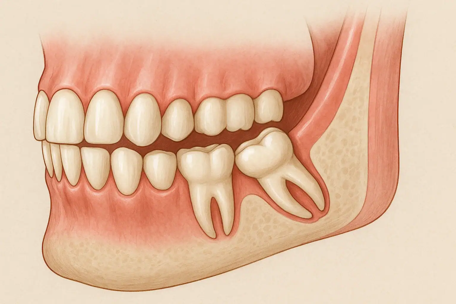
Radiographs classify an impacted wisdom tooth by its angulation. A mesioangular tooth tilts forward toward the front of the mouth, a vertical tooth is upright but stuck, a distoangular tooth tilts backward, and a horizontal tooth lies on its side within the jaw. These angles are not evenly spread. In the meta-analysis of 49 studies, mesioangular impaction was the most common at 41.17%, followed by vertical at 25.55%, distoangular at 12.17% and horizontal at 11.06% (PubMed 26561441).
Angulation matters because it shapes how the tooth is approached surgically. A mesioangular tooth often presses against the back of the second molar, and a horizontal tooth usually needs to be sectioned before removal. Depth in the bone and the distance to the nerve are also read from the imaging, and together these findings decide whether the tooth is monitored, removed in full, or managed with a more conservative approach.
Treatment by Monitoring, Removal or Coronectomy
Management runs from radiographic monitoring to surgical removal, and the choice depends on what the impacted tooth is doing. Asymptomatic impacted wisdom teeth that are not harming their neighbours may be kept under routine observation with periodic radiographs.
Removal is considered when there is decay in the neighbouring tooth, recurrent gum infection around the tooth, a cyst, damage to adjacent roots, or crowding pressure. Surgical extraction is performed under local anaesthesia, and sedation or general anaesthesia can be used for more complex cases or anxious patients.
When a lower wisdom tooth lies very close to the inferior alveolar nerve, coronectomy is an alternative to full removal. In this procedure the crown is removed and the roots are left in place, away from the nerve. A meta-analysis of 34 studies covering 7,115 wisdom teeth in 4,477 patients found that coronectomy lowered the risk of inferior alveolar nerve injury compared with extraction (RR 0.1, 95% CI 0.0 to 0.2) (PubMed 39956152). It also reduced lingual nerve injury and dry socket, with no significant difference in postoperative infection (PubMed 39956152).
Coronectomy has its own trade-offs. The same analysis reported a failure rate of 2.79% (119 of 4,261), surgical reintervention in 3.63% (111 of 3,055) and root exposure in 2.66% (61 of 2,290) (PubMed 39956152). It is therefore reserved for teeth where the nerve risk of full extraction is high.
Impacted Canines and Early Orthodontic Checks
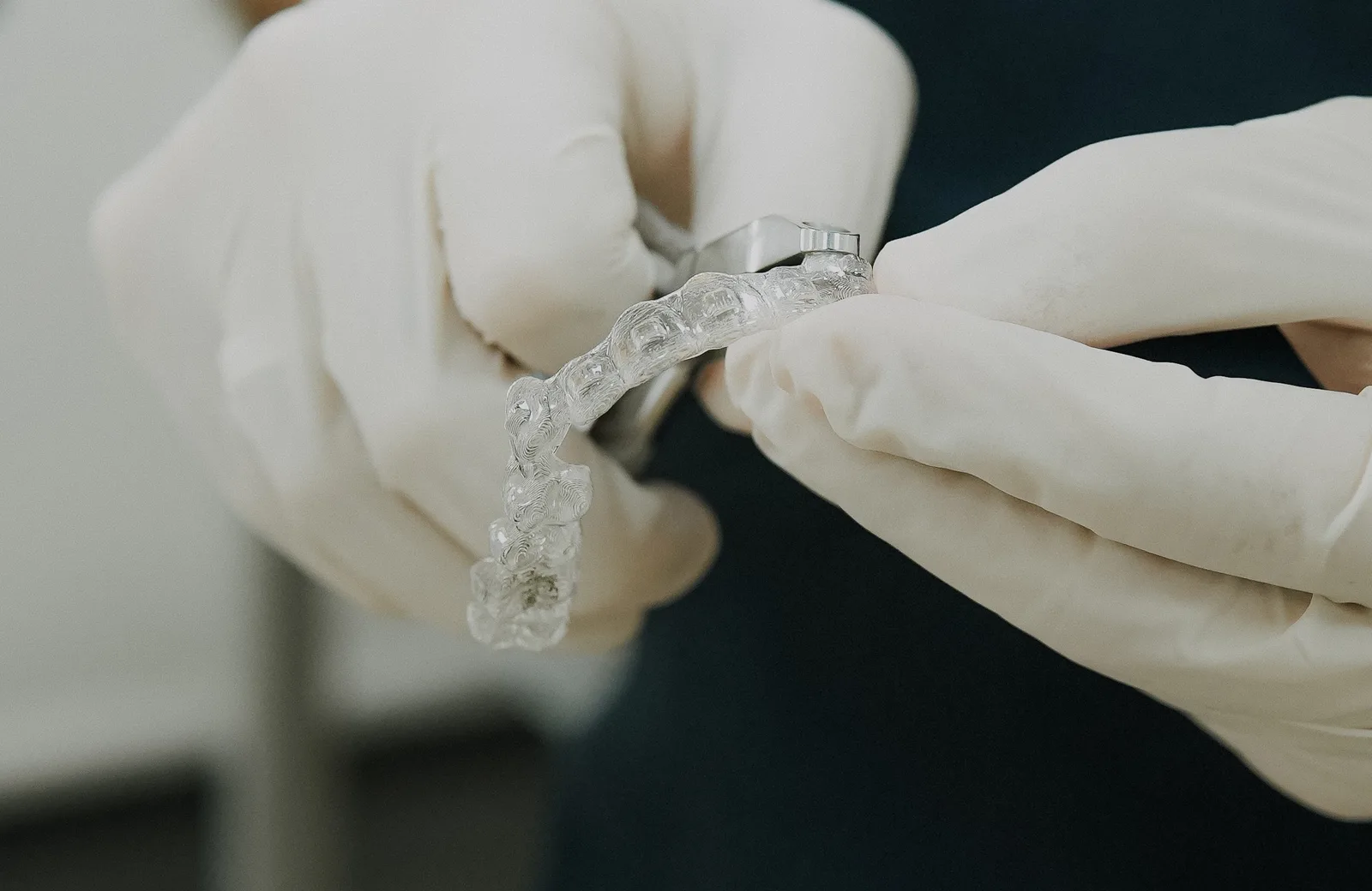
Impacted canines are usually not extracted, because the canine anchors the corner of the smile and guides the bite when the jaw moves sideways. The standard route is exposure and bracketing, carried out together with orthodontic treatment. The tooth is surgically uncovered, a small bracket with a chain is bonded to it, and gentle orthodontic traction pulls it into the arch over months.
Before the canine is moved, the orthodontist usually creates space for it in the arch with braces, so that the tooth has somewhere to go. The traction is slow and controlled to protect the roots of the neighbouring incisors, which an impacted canine can press on. A deeply positioned canine takes longer to bring down.
The caution around the incisor roots has a measured basis. A systematic review of 18 cone beam CT studies found that impacted upper canines sit on the palatal side in 56.99% of cases; among the resorbed roots of neighbouring teeth, resorption was slight in 43.2% and severe in 30.9%, and the apical third of the root was affected most often (PubMed 30165975). The authors link the resorption to contact between the erupting canine and the incisor roots and note that delayed eruption or delayed treatment may lead to resorption of the lateral and central incisors (PubMed 30165975), which is why a canine found early leaves more options.
Early orthodontic monitoring, typically around ages 7 to 10, is what makes this route easier. At that age a dentist or orthodontist can check whether the canines are developing on a normal path and whether the baby canines are loosening on time.
Recovery After Surgery and Follow-Up
Recovery after removal or exposure of an impacted tooth usually involves mild swelling and soreness for a few days. Ice packs on the cheek reduce swelling in the first day. A soft diet is kept for 2 to 3 days, and salt water rinses start after 24 hours to keep the area clean without disturbing it.
The blood clot that forms in the socket protects the bone and is the base for healing. Smoking, drinking through a straw and vigorous rinsing are avoided in the first days because they can dislodge the clot, which can lead to dry socket, a painful condition in which the bone is left exposed. Pain relief follows the surgeon's instructions.
Pain that increases instead of easing, swelling that keeps growing, fever, pus, or numbness of the lip or tongue that persists after the anaesthesia has worn off should be reported without waiting for the scheduled visit.
A follow-up appointment confirms that the area is healing and rules out dry socket or infection. After coronectomy, follow-up radiographs check the position of the retained roots over time. After canine exposure, the orthodontic visits continue until the tooth has reached its place in the arch.
Let’s plan the right treatment together.
Free Assessment→- 1.Carter K, Worthington S. Predictors of Third Molar Impaction: A Systematic Review and Meta-analysis. J Dent Res 2016;95:267-76. PubMed 26561441
- 2.Kang FW, Yuan XR, Li GC, Yang YFZ, Zhang XM, Hou GY. Coronectomy in Lower Third Molar Surgery: A Systematic Review and Meta-Analysis. J Oral Maxillofac Surg 2025;83:601-615. PubMed 39956152
- 3.Pinto AC, Francisco H, Marques D, Martins JNR, Caramês J. Worldwide Prevalence and Demographic Predictors of Impacted Third Molars-Systematic Review with Meta-Analysis. J Clin Med 2024;13:7533. PubMed 39768456
- 4.Schroder AGD, Guariza-Filho O, de Araujo CM, Ruellas AC, Tanaka OM, Porporatti AL. To what extent are impacted canines associated with root resorption of the adjacent tooth?: A systematic review with meta-analysis. J Am Dent Assoc 2018;149:765-777.e8. PubMed 30165975