Digital dentistry is the delivery of dental care through computer-aided systems, in which the impression, the diagnosis, the treatment plan and the fabrication all run on a shared three-dimensional data set instead of trays, stone models and manual waxing. Its accuracy is measurable rather than absolute: in an in vivo study of 11 participants scanned with three intraoral scanners, median trueness was 38 to 47 micrometres for short-span scans and 147 to 433 micrometres for complete-arch scans (PubMed 33902891). The working tools are intraoral scanners, CAD/CAM design and milling, 3D printing and digital radiography, and the practical gain is fewer appointments and same-visit restorations.
- 01Digital dentistry replaces the analogue chain of tray impression, stone model and hand fabrication with a scan, design and manufacture chain built on one 3D file.
- 02Scanner accuracy depends on the span being scanned: median in vivo trueness was 38 to 47 micrometres for short-span scans against 147 to 433 micrometres for complete-arch scans, across three devices and 11 participants (PubMed 33902891).
- 03In the same study, extraoral scanning of the model was more precise and truer than intraoral scanning for almost every comparison, so the mouth itself is the limiting condition, not the software (PubMed 33902891).
- 04Full-arch implant scanning is a separate measurement problem: resultant trueness was 41 plus or minus 11 micrometres with the control protocol, 49 plus or minus 22 with dental floss and 55 plus or minus 8 with an acrylic splint (PubMed 36481688).
- 05CAD/CAM shortens the laboratory step rather than the clinical one: crowns, onlays, veneers and temporary restorations can be designed and milled in hours, which removes shipping and model pouring from the timeline.
/ Digital Dentistry Definition, Evolution and 3D Mouth Scanners
Digital dentistry is a modern treatment approach that is formed by transforming classical dentistry methods with computer-aided systems. Thanks to this technology, all processes from taking measurements to diagnosis, from treatment planning to application are carried out on digital platforms.
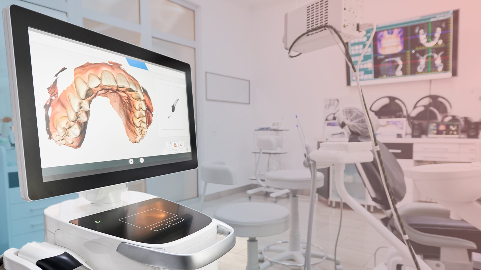
Prosthesis preparations that can take weeks in traditional methods can be completed in a few hours. Silicone trays used during measurement have given way to 3D mouth scanners. Prostheses are prepared in a digital environment with CAD/CAM systems and produced with 3D printers.
In short: The era has changed in dentistry and the name of this era is "digitalization".
History and Evolution of Digital Dentistry
Digital dentistry began with the first use of CAD/CAM systems in the 1980s. These systems, first used in fixed prosthesis design, became widespread in the early 2000s.
Today, we have:
- Digital measurement systems
- Three-dimensional printers
- AI-supported diagnostic software
- Digital tomography systems
- In-clinic laboratory automations
the cornerstones of digital dentistry
3D Mouth Scanners
Traditional impression taking methods such as impression trays and silicone materials negatively affect patient comfort. 3D mouth scanners fundamentally change this process:
- The inside of the mouth is scanned in seconds.
- A 3D model is created in a digital environment.
- It works with a precision of one thousandth of a millimeter.
- Analysis and recording can be done simultaneously.
This system is indispensable especially for patients with gag reflexes.
/ CAD/CAM, Imaging and Patient Experience in Digital Dentistry
CAD/CAM Systems
CAD/CAM (Computer-Aided Design / Computer-Aided Manufacturing) systems enable the design of prostheses in a digital environment and production on the same day.
- With this technology:
- Restorations such as crowns, bridges, veneers
- Onlay/inlay fillings
- Temporary prostheses
can be produced in hours. Errors are minimized, there is no need for repetition.
Artificial Intelligence-Assisted Diagnostic Systems
Artificial intelligence dentistry strengthens the diagnostic process. X-ray images are scanned with artificial intelligence algorithms.
The system analyzes caries, root canal needs, gum disease and bone density.
This way:
- Diagnosis time is shortened
- Doctor error is reduced
- Decision processes are accelerated
Digital X-Ray and Imaging
Digital X-Rays contain up to 70% less radiation than traditional X-Rays.
In addition:
- Instant results are obtained
- Images are recorded digitally
- Details are analyzed with zoom and filtering
- Can be easily stored in the patient file
Prosthesis Production with 3D Printers
Thanks to 3D printers:
- Night guards
- Temporary teeth
- Orthodontic appliances
- Surgical guides
can be produced directly from digital models. This eliminates laboratory time.
The Impact of Digital Dentistry on Patient Experience
The benefits of digital systems for patients are:
- Procedures completed in a short time
- Fewer appointments
- Less pain
- Comfortable measurement
- Visual presentation of the treatment process
- Preview of aesthetic results
Especially in aesthetic applications such as smile design, the patient can see their new smile before the procedure.
Advantages of Digitalization from a Dentist Perspective
- More precise measurement
- Shorter treatment time
- Reduced risk of manual errors
- Digital archiving of patient records
- Time savings with in-clinic production
- Increased patient satisfaction
Especially in clinics with high patient traffic, digital systems greatly increase efficiency.
/ Application Areas, Education and Data Security in Digital Dentistry
Digital Smile Design
A smile simulation is prepared in a digital environment by taking into account the person's:
- Facial structure
- Smile line
- Tooth-lip ratio
with face recognition and analysis software.
Implantology
Digital planning in implant applications:
- Angle where the implant will be placed
- Depth
- Jawbone density
is analyzed in advance. The margin of error is reduced to a minimum with surgical guides.
Orthodontics
Clear aligner treatments are carried out completely with digital planning.
The patient can watch the entire process in 3D before using the aligners. This increases motivation.
Pedodontics
The traumatic effect is eliminated by digitalizing the measurement process in child patients. At the same time, fears are reduced with gamified applications.
Endodontics and Restorative Treatments
With canal length measurements and CAD-supported fillings:
- Shorter root canal treatment
- More durable filling materials
- More aesthetic results are achieved
Education and the Future in Digital Dentistry
Today's dentistry students now receive simulation training using 3D software.
They plan restorations using digital patient models, not real patients.
Foresight for the future:
- Robotic tooth extraction
- Remote-controlled devices
- Fully automated prosthesis production
- Mobile intraoral scanners
Data Security in Digital Applications
Since patient information is stored in a digital environment, data security is of critical importance.
For this purpose:
- SSL certified systems
- Encrypted data access
- Cloud backup systems
- Transaction approvals with e-signature
are used. Patient privacy is maximized with KVKK-compliant archiving.
/ Frequently Asked Questions
Is digital dentistry safe?
Yes. All systems are CE and FDA approved. Fully compliant with health standards.
How precise are digital measurements?
Measurements can be made in a thousandth of a millimeter. It is much more precise than classical methods.
Is a same-day prosthesis possible?
Yes. Thanks to CAD/CAM technology, the entire process from scanning the tooth to applying the prosthesis can be completed in a few hours.
Is digital x-ray harmful?
No. It contains much less radiation than traditional x-rays. Maximum safety is provided with advanced devices. For scale, a phantom study measured 14.2 to 24.3 microsieverts for a digital panoramic film, against 388 microsieverts for a full set of intraoral films on older D-speed film with round collimation, and it lists digital receptors among the main ways to reduce patient dose. For scale, a phantom study measured 14.2 to 24.3 microsieverts for a digital panoramic film, against 388 microsieverts for a full set of intraoral films on older D-speed film with round collimation, and it lists digital receptors among the main ways to reduce patient dose.
The System That Brings the Future to Life Today: Digital Dentistry
Digital dentistry is not a technology choice; it is a requirement of the age. It offers a more accurate, faster, more comfortable and more reliable system for both the doctor and the patient.
The treatment process has been reorganized to work with digital systems in every field, from smile design to implantology, from orthodontics to pedodontics.
You do not have to wait for the future. Because digital dentistry carries the future.
Let’s plan the right treatment together.
Free Assessment→- 1.Kernen F, Schlager S, Seidel Alvarez V, Mehrhof J, Vach K, Kohal R, et al. Accuracy of intraoral scans: An in vivo study of different scanning devices. J Prosthet Dent 2022;128:1303-1309. PubMed 33902891
- 2.Nedelcu R, Olsson P, Thulin M, Nyström I, Thor A. In vivo trueness and precision of full-arch implant scans using intraoral scanners with three different acquisition protocols. J Dent 2023;128:104308. PubMed 36481688
- 3.Ludlow JB, Davies-Ludlow LE, White SC. Patient risk related to common dental radiographic examinations: the impact of 2007 International Commission on Radiological Protection recommendations regarding dose calculation. J Am Dent Assoc. 2008;139(9):1237-43.